 Explore
 Validate
 Learn
Learn Western blot
Antibody data
- Antibody Data
- Antigen structure
- References [4]
- Comments [0]
- Validations
- Western blot [5]
- Immunocytochemistry [2]
- Immunohistochemistry [3]
- Other assay [3]
Submit
Validation data
Reference
Comment
Report error
- Product number
- MA1-117 - Provider product page

- Provider
- Invitrogen Antibodies
- Product name
- PAX8 Monoclonal Antibody (1F8-3A8)
- Antibody type
- Monoclonal
- Antigen
- Purifed from natural sources
- Description
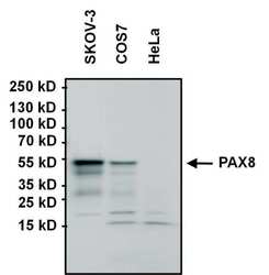
- Western blot analysis of MA1-117 detects a prominent ~48 kDa protein in human adenocarcinoma SKOV-3 and SV40 transformed African green monkey (Cercopithecus aethiops) kidney cells. In SKOV-3 cells, additional unknown low molecular bands are also detected. Specificity of the antibody was confirmed in HeLa (negative control) and 293T cells overexpressing full length PAX8.
- Reactivity
- Human, Mouse
- Host
- Mouse
- Isotype
- IgG
- Antibody clone number
- 1F8-3A8
- Vial size
- 100 µg
- Concentration
- 1 mg/mL
- Storage
- -20°C
Submitted references A Stem Cell Surge During Thyroid Regeneration.
Derivation and 97% Purification of Human Thyroid Cells From Dermal Fibroblasts.
The Repertoire of Serous Ovarian Cancer Non-genetic Heterogeneity Revealed by Single-Cell Sequencing of Normal Fallopian Tube Epithelial Cells.
Acute renal failure due to small cell neuroendocrine carcinoma of the left kidney: A case report.
Ma R, Morshed SA, Latif R, Davies TF
Frontiers in endocrinology 2020;11:606269
Frontiers in endocrinology 2020;11:606269
Derivation and 97% Purification of Human Thyroid Cells From Dermal Fibroblasts.
Ma R, Shi R, Morshed SA, Latif R, Davies TF
Frontiers in endocrinology 2020;11:446
Frontiers in endocrinology 2020;11:446
The Repertoire of Serous Ovarian Cancer Non-genetic Heterogeneity Revealed by Single-Cell Sequencing of Normal Fallopian Tube Epithelial Cells.
Hu Z, Artibani M, Alsaadi A, Wietek N, Morotti M, Shi T, Zhong Z, Santana Gonzalez L, El-Sahhar S, Carrami EM, Mallett G, Feng Y, Masuda K, Zheng Y, Chong K, Damato S, Dhar S, Campo L, Garruto Campanile R, Soleymani Majd H, Rai V, Maldonado-Perez D, Jones S, Cerundolo V, Sauka-Spengler T, Yau C, Ahmed AA
Cancer cell 2020 Feb 10;37(2):226-242.e7
Cancer cell 2020 Feb 10;37(2):226-242.e7
Acute renal failure due to small cell neuroendocrine carcinoma of the left kidney: A case report.
Haberal HB, Tonyali Ş, Baydar DE, Bilen CY
Oncology letters 2017 Nov;14(5):6117-6120
Oncology letters 2017 Nov;14(5):6117-6120
No comments: Submit comment
Supportive validation
- Submitted by
- Invitrogen Antibodies (provider)
- Main image
- Experimental details
- Western blot analysis of PAX8 was performed by loading 60 µg of SKOV-3, COS7, and negative control HeLa whole cell lysates, and 10 µL of PageRuler Plus Prestained Protein Ladder (Product # 26619) onto a 4-20% Tris-HCl polyacrylamide gel. Proteins were transferred to a PVDF membrane and blocked with 5% BSA/TBST for at least 1 hour. The membrane was probed with a PAX8 monoclonal antibody (Product # MA1-117) at a dilution of 1:1000 overnight at 4°C on a rocking platform, washed in TBS-0.1%Tween-20, and probed with a pre-diluted (10 µg/mL) HRP-conjugated goat anti-mouse IgG secondary antibody (Product # 32430) at a dilution of 1:500 for 1 hour. Chemiluminescent detection was performed using SuperSignal West Pico (Product # 34080).

- Submitted by
- Invitrogen Antibodies (provider)
- Main image

- Experimental details
- Western blot analysis of PAX8 was performed by loading 25 µg of HEK293T cell lysate overexpressing PAX8 (right lane) or empty vector control (left lane), and 10 µL PageRuler Plus Prestained Protein Ladder (Product # 26619) onto a 4-20% Tris-HCl polyacrylamide gel. Proteins were transferred to a PVDF membrane and blocked with 5% BSA/TBST for at least 1 hour. The membrane was probed with a PAX8 monoclonal antibody (Product # MA1-117) at a dilution of 1:1000 overnight at 4°C on a rocking platform, washed in TBS-0.1%Tween-20, and probed with a pre-diluted (10 µg/mL) HRP-conjugated goat anti-mouse IgG secondary antibody (Product # 32430) at a dilution of 1:500 for 1 hour. Chemiluminescent detection was performed using SuperSignal West Pico (Product # 34080).

- Submitted by
- Invitrogen Antibodies (provider)
- Main image

- Experimental details
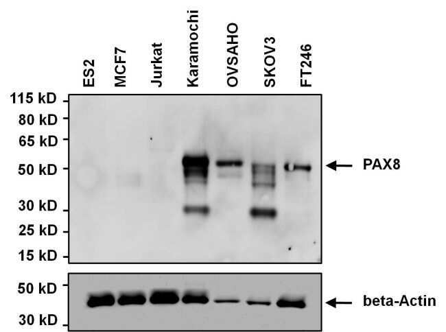
- Western blot analysis of PAX8 was performed by loading 20 µg of the indicated whole cell lysates onto a 4-12% Novex Bis-Tris polyacrylamide gel. Proteins were transferred to a nitrocellulose membrane and blocked with 5% milk in PBST for 1 hour. The membrane was probed with a PAX8 monoclonal antibody (Product # MA1-117) at a dilution of 1:500 overnight at 4C on a rocking platform, washed in PBST, and probed with a goat anti-mouse IgG secondary antibody. Samples were also probed with a beta-actin antibody to control for equal protein loading. Chemiluminescent detection was performed using ECL Plus substrate (Product # 32132). The lysates were: ES2 ovarian clear cell carcinoma, MCF7 breast cancer, Jurkat T cell leukemia, Kuramochi high grade serious ovarian cancer, OVSAHO high grade serious ovarian carcinoma, SKOV3 ovarian adenocarcinoma, and FT246 immortalized fallopian tube epithelium cells. Data courtesy of the Innovators Program.
- Submitted by
- Invitrogen Antibodies (provider)
- Main image

- Experimental details
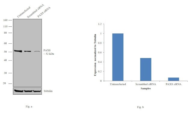
- Knockdown of PAX8 was achieved by transfecting COS-7 cells with PAX8 specific siRNAs (Silencer® select Product # n304366, s15404). Western blot analysis (Fig. a) was performed using whole cell extracts from the PAX8 knockdown cells (lane 3), non-specific scrambled siRNA transfected cells (lane 2) and untransfected cells (lane 1). The blots were probed with Anti-PAX8 Monoclonal Antibody (Product # MA1-117, 1:1000 dilution) and Goat anti-Mouse IgG (H+L) Superclonal™ Secondary Antibody, HRP conjugate (Product # A28177, 0.25 µg/mL, 1:4000 dilution). Densitometric analysis of this western blot is shown in histogram (Fig. b). Decrease in signal upon siRNA mediated knock down confirms that antibody is specific to PAX8.
- Submitted by
- Invitrogen Antibodies (provider)
- Main image

- Experimental details
- Western blot analysis was performed on modified whole cell extracts (1% SDS) (30 µg lysate) of COS-7 (Lane 1), HEK-293 (Lane 2), HeLa (Lane 3), Jurkat (Lane 4) and MCF7 (Lane 5). The blot was probed with Anti-PAX8 Monoclonal Antibody (1F8-3A8) (Product # MA1-117, 1:500 dilution) and detected by chemiluminescence using Goat anti-Mouse IgG (H+L) Superclonal™ Secondary Antibody, HRP conjugate (Product # A28177, 0.25 µg/mL, 1:4000 dilution). A 50 kDa band corresponding to PAX8 was detected in COS-7 and not in HEK-293, HeLa, Jurkat and MCF7 which are reported negative for PAX8. An uncharacterized band was observed ~35 kDa across cell lines tested.

Supportive validation
- Submitted by
- Invitrogen Antibodies (provider)
- Main image

- Experimental details
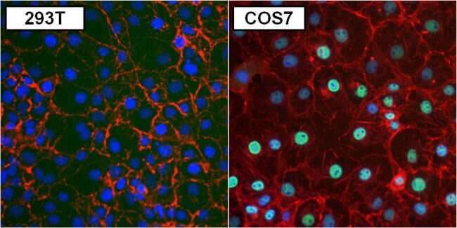
- Immunofluorescent analysis of PAX8 (green) in 293T (left panel) and COS7 cells (right panel). Formalin fixed cells were permeabilized with 0.1% Triton X-100 in TBS for 10 minutes at room temperature. Cells were blocked with 1% Blocker BSA (Product # 37525) for 15 minutes at room temperature. Cells were probed with a PAX8 monoclonal antibody (Product # MA1-117) at a dilution of 1:50 for at least 1 hour at room temperature, washed with PBS, and incubated with a DyLight 488-conjugated goat anti-mouse IgG secondary antibody (Product # 35502) at a dilution of 1:400 for 30 minutes at room temperature. F-Actin (red) was stained with DyLight-554 Phalloidin (Product # 21834) and nuclei (blue) were stained with Hoechst 33342 dye (Product # 62249). Images were taken on a Thermo Scientific ToxInsight Instrument at 20X magnification.
- Submitted by
- Invitrogen Antibodies (provider)
- Main image
- Experimental details
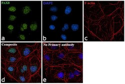
- Immunofluorescence analysis of PAX8 was performed using 70% confluent log phase COS-7 cells. The cells were fixed with 4% paraformaldehyde for 10 minutes, permeabilized with 0.1% Triton™ X-100 for 15 minutes, and blocked with 1% BSA for 1 hour at room temperature. The cells were labeled with PAX8 Monoclonal Antibody (1F8-3A8) (Product # MA1-117) at 1:100 dilution in 0.1% BSA, incubated at 4 degree Celsius overnight and then labeled with Goat anti-Mouse IgG (H+L) Superclonal™ Secondary Antibody, Alexa Fluor® 488 conjugate (Product #A28175) at a dilution of 1:2000 for 45 minutes at room temperature (Panel a: green). Nuclei (Panel b: blue) were stained with ProLong™ Diamond Antifade Mountant with DAPI (Product # P36962). F-actin (Panel c: red) was stained with Rhodamine Phalloidin (Product # R415, 1:300). Panel d represents the merged image showing Nuclear localization. Panel e represents control cells with no primary antibody to assess background. The images were captured at 60X magnification.

Supportive validation
- Submitted by
- Invitrogen Antibodies (provider)
- Main image
- Experimental details
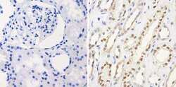
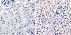
- Immunohistochemistry analysis of PAX8 showing staining in the nucleus of paraffin-embedded human kidney tissue (right) compared to a negative control without primary antibody (left). To expose target proteins, antigen retrieval was performed using 10mM sodium citrate (pH 6.0), microwaved for 8-15 min. Following antigen retrieval, tissues were blocked in 3% H2O2-methanol for 15 min at room temperature, washed with ddH2O and PBS, and then probed with a PAX8 monoclonal antibody (Product # MA1-117) diluted in 3% BSA-PBS at a dilution of 1:100 overnight at 4°C in a humidified chamber. Tissues were washed extensively in PBST and detection was performed using an HRP-conjugated secondary antibody followed by colorimetric detection using a DAB kit. Tissues were counterstained with hematoxylin and dehydrated with ethanol and xylene to prep for mounting.
- Submitted by
- Invitrogen Antibodies (provider)
- Main image

- Experimental details
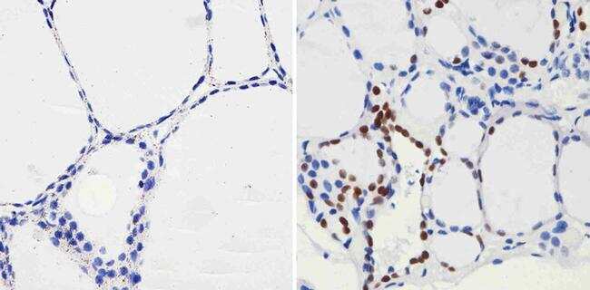
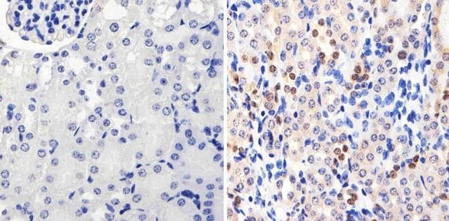
- Immunohistochemistry analysis of PAX8 showing staining in the nucleus of paraffin-embedded human thyroid tissue (right) compared to a negative control without primary antibody (left). To expose target proteins, antigen retrieval was performed using 10mM sodium citrate (pH 6.0), microwaved for 8-15 min. Following antigen retrieval, tissues were blocked in 3% H2O2-methanol for 15 min at room temperature, washed with ddH2O and PBS, and then probed with a PAX8 monoclonal antibody (Product # MA1-117) diluted in 3% BSA-PBS at a dilution of 1:100 overnight at 4°C in a humidified chamber. Tissues were washed extensively in PBST and detection was performed using an HRP-conjugated secondary antibody followed by colorimetric detection using a DAB kit. Tissues were counterstained with hematoxylin and dehydrated with ethanol and xylene to prep for mounting.
- Submitted by
- Invitrogen Antibodies (provider)
- Main image
- Experimental details
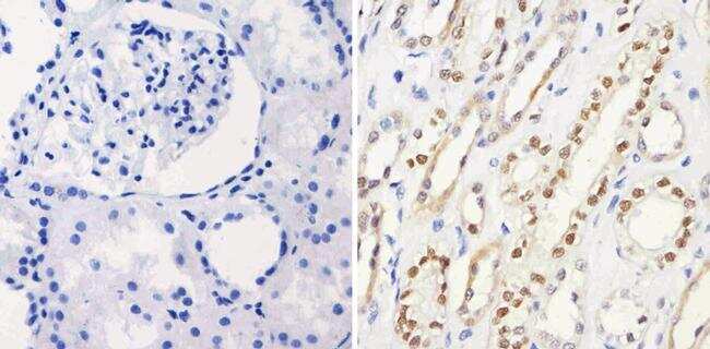
- Immunohistochemistry analysis of PAX8 showing staining in the nucleus of paraffin-embedded mouse kidney tissue (right) compared to a negative control without primary antibody (left). To expose target proteins, antigen retrieval was performed using 10mM sodium citrate (pH 6.0), microwaved for 8-15 min. Following antigen retrieval, tissues were blocked in 3% H2O2-methanol for 15 min at room temperature, washed with ddH2O and PBS, and then probed with a PAX8 monoclonal antibody (Product # MA1-117) diluted in 3% BSA-PBS at a dilution of 1:20 overnight at 4°C in a humidified chamber. Tissues were washed extensively in PBST and detection was performed using an HRP-conjugated secondary antibody followed by colorimetric detection using a DAB kit. Tissues were counterstained with hematoxylin and dehydrated with ethanol and xylene to prep for mounting.
Supportive validation
- Submitted by
- Invitrogen Antibodies (provider)
- Main image

- Experimental details
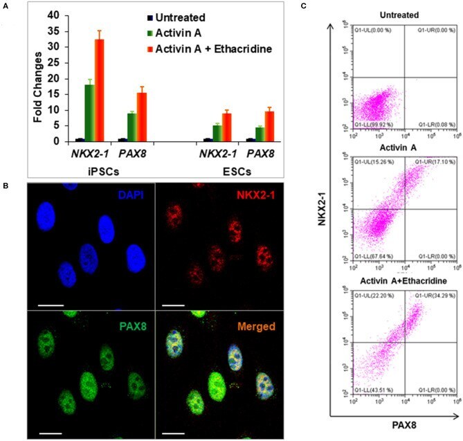
- Figure 4 Differentiation of definitive thyroid endoderm from human iPSCs (LF cells). (A) qPCR analysis of thyroid transcriptional factors NKX2-1 and PAX8 in human ES cells and human iPSCs (LF cells). Fold change is represented as the mean +- SEM of three independent experiments. (B) Immunostaining of NKX2-1 (Red) and PAX8 (Green) in human iPSCs (LF cells) derived thyroid endoderm cells. Scale bar = 20 mum. (C) Representative flow cytometry of NKX2-1 and PAX8 expressing cells in untreated (upper), activin A treated (middle) and activing A + Ethacridine (lower) cells. Data were representative of three separate experiments.
- Submitted by
- Invitrogen Antibodies (provider)
- Main image

- Experimental details
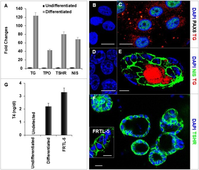
- Figure 6 Characterization of differentiated thyroid cells. (A) qPCR analysis of thyroid specific genes: TG, TPO, TSHR, and NIS. Fold change is represented as the mean +- SEM of three independent experiments on differentiated LF cells at 21 days. (B-F) Immunostaining of thyroid genes in undifferentiated and differentiated LF cells at 21 days culture: (B,D) Undifferentiated cells as a control. (C) Staining of TG (Red) and PAX8 (Green) in differentiated cells. TG expressed in cytoplasm and PAX8 expressed in nucleus. (E) Staining of TG (Red) and NIS (Green) in a differentiated thyroid follicle: NIS was expressed in the membrane and TG was expressed in the cytoplasm and follicular lumen. (F) Thyroid neo-follicles derived from differentiated cells expressing TSHR (Green) in the membrane. Inset shows staining of rat FRTL-5 thyroid cells. Scale bar = 20 mum. (G) Measurement of T4 from the differentiated LF cells: T4 was detected in the iodine supplemented medium of the differentiated LF cells at 23 days culture as described in Methods and was absent in the medium of the undifferentiated cells but less than in FRTL-5 cells with 7H medium.
- Submitted by
- Invitrogen Antibodies (provider)
- Main image
- Experimental details
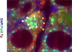
- Figure 7 Representative images of immunostaining for expression of Tg (red), Pax8 (green), and DAPI (blue) in the thyroid gland of TPOCreER2/iDTR mice injected with TM/DT for 2 weeks. Note the larger parafollicular collections of cells positive for Pax8 (green) and negative for Tg (labeled with white arrows) indicative of progenitor cells and Pax 8 positive and Tg positive cells (labelled with orange arrows) indicative of more mature cells. There are also Pax8 and Tg negative cells (blue) which are likely fibroblast or other cell types.
