Explore
 Validate
 Learn
Learn Western blot
 Immunocytochemistry
Antibody data
- Antibody Data
- Antigen structure
- References [1]
- Comments [0]
- Validations
- Western blot [3]
- Immunohistochemistry [1]
Submit
Validation data
Reference
Comment
Report error
- Product number
- NBP1-89818 - Provider product page

- Provider
- Novus Biologicals
- Proper citation
- Novus Cat#NBP1-89818, RRID:AB_11003647
- Product name
- Rabbit Polyclonal Alpha Actinin 4 Antibody
- Antibody type
- Polyclonal
- Description
- Immunogen affinity purified. Specificity of human, mouse, rat Alpha Actinin 4 antibody verified on a Protein Array containing target protein plus 383 other non-specific proteins.
- Reactivity
- Human, Mouse, Rat
- Host
- Rabbit
- Isotype
- IgG
- Vial size
- 0.1 ml
- Storage
- Store at 4C short term. Aliquot and store at -20C long term. Avoid freeze-thaw cycles.
Submitted references Systematic validation of antibody binding and protein subcellular localization using siRNA and confocal microscopy.
Stadler C, Hjelmare M, Neumann B, Jonasson K, Pepperkok R, Uhlén M, Lundberg E
Journal of proteomics 2012 Apr 3;75(7):2236-51
Journal of proteomics 2012 Apr 3;75(7):2236-51
No comments: Submit comment
Supportive validation
- Submitted by
- Novus Biologicals (provider)
- Main image

- Experimental details
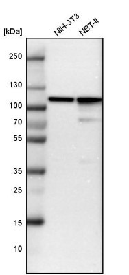
- Western Blot: Alpha Actinin 4 Antibody [NBP1-89818] - Analysis in mouse cell line NIH-3T3 and rat cell line NBT-II.
- Submitted by
- Novus Biologicals (provider)
- Main image
- Experimental details
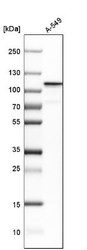
- Western Blot: Alpha Actinin 4 Antibody [NBP1-89818] - Analysis in human cell line A-549.

- Submitted by
- Novus Biologicals (provider)
- Main image

- Experimental details
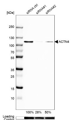
- Western Blot: Alpha Actinin 4 Antibody [NBP1-89818] - Analysis in A-549 cells transfected with control siRNA, target specific siRNA probe #1 and #2,. Remaining relative intensity is presented. Loading control: Anti-GAPDH.
Supportive validation
- Submitted by
- Novus Biologicals (provider)
- Main image

- Experimental details
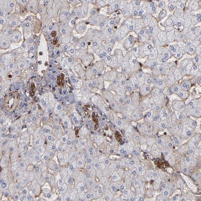
- Immunohistochemistry-Paraffin: Alpha Actinin 4 Antibody [NBP1-89818] - Staining of human liver shows strong cytoplasmic and membranous positivity in bile duct cells.