 Explore
 Validate
 Learn
Learn Flow cytometry
Antibody data
- Antibody Data
- Antigen structure
- References [5]
- Comments [0]
- Validations
- Flow cytometry [1]
Submit
Validation data
Reference
Comment
Report error
- Product number
- 48-0388-41 - Provider product page

- Provider
- Invitrogen Antibodies
- Product name
- Anti-CD38 Monoclonal Antibody (HB7), eFluor 450, eBioscience™
- Antibody type
- Monoclonal
- Antigen
- Other
- Description
- Description: The HB7 monoclonal antibody reacts with the human CD38 molecule, an approximately 45 kDa type II transmembrane protein. CD38 is an ectoenzyme which catalyses NAD into nicotinic acid adenine dinucleotide phosphate (NAADP) and cyclic ADP-ribose (cADPR), both of which are secondary messengers. Expression of CD38 is bimodal during B cell development, modulating from high in immature cells to low in intermediate ones and back to high on mature B cells. Additionally CD38 is found in a variety of tissues and other hematopoietic cells (e.g. T cells, NK cells and monocytes) and can be used to phenotype leukemias and monitor HIV-1 progression. The CD34+CD38- population of hematopoietic stems cells is thought to define the most pluripotent cells (e.g. blast colony forming cells). In addition to surface expression, CD38 has recently been found in the nucleus where it may play a role in monitoring calcium levels. Applications Reported: This HB7 antibody has been reported for use in flow cytometric analysis. Applications Tested: This HB7 antibody has been pre-titrated and tested by flow cytometric analysis of normal human peripheral blood cells. This can be used at 5 µL (0.06 µg) per test. A test is defined as the amount (µg) of antibody that will stain a cell sample in a final volume of 100 µL. Cell number should be determined empirically but can range from 10^5 to 10^8 cells/test. eFluor® 450 is an alternative to Pacific Blue®. eFluor® 450 emits at 445 nm and is excited with the Violet laser (405 nm). Please make sure that your instrument is capable of detecting this fluorochome. Excitation: 405 nm; Emission: 445 nm; Laser: Violet Laser. Filtration: 0.2 µm post-manufacturing filtered.
- Reactivity
- Human
- Host
- Mouse
- Isotype
- IgG
- Antibody clone number
- HB7
- Vial size
- 25 Tests
- Concentration
- 5 µL/Test
- Storage
- 4° C, store in dark, DO NOT FREEZE!
Submitted references UTX/KDM6A Loss Enhances the Malignant Phenotype of Multiple Myeloma and Sensitizes Cells to EZH2 inhibition.
CD38 is constitutively expressed in the nucleus of human hematopoietic cells.
Anti-CD38-blocked ricin: an immunotoxin for the treatment of multiple myeloma.
Sequential generations of hematopoietic colonies derived from single nonlineage-committed CD34+CD38- progenitor cells.
Isolation of a cDNA encoding the human CD38 (T10) molecule, a cell surface glycoprotein with an unusual discontinuous pattern of expression during lymphocyte differentiation.
Ezponda T, Dupéré-Richer D, Will CM, Small EC, Varghese N, Patel T, Nabet B, Popovic R, Oyer J, Bulic M, Zheng Y, Huang X, Shah MY, Maji S, Riva A, Occhionorelli M, Tonon G, Kelleher N, Keats J, Licht JD
Cell reports 2017 Oct 17;21(3):628-640
Cell reports 2017 Oct 17;21(3):628-640
CD38 is constitutively expressed in the nucleus of human hematopoietic cells.
Orciani M, Trubiani O, Guarnieri S, Ferrero E, Di Primio R
Journal of cellular biochemistry 2008 Oct 15;105(3):905-12
Journal of cellular biochemistry 2008 Oct 15;105(3):905-12
Anti-CD38-blocked ricin: an immunotoxin for the treatment of multiple myeloma.
Goldmacher VS, Bourret LA, Levine BA, Rasmussen RA, Pourshadi M, Lambert JM, Anderson KC
Blood 1994 Nov 1;84(9):3017-25
Blood 1994 Nov 1;84(9):3017-25
Sequential generations of hematopoietic colonies derived from single nonlineage-committed CD34+CD38- progenitor cells.
Terstappen LW, Huang S, Safford M, Lansdorp PM, Loken MR
Blood 1991 Mar 15;77(6):1218-27
Blood 1991 Mar 15;77(6):1218-27
Isolation of a cDNA encoding the human CD38 (T10) molecule, a cell surface glycoprotein with an unusual discontinuous pattern of expression during lymphocyte differentiation.
Jackson DG, Bell JI
Journal of immunology (Baltimore, Md. : 1950) 1990 Apr 1;144(7):2811-5
Journal of immunology (Baltimore, Md. : 1950) 1990 Apr 1;144(7):2811-5
No comments: Submit comment
Supportive validation
- Submitted by
- Invitrogen Antibodies (provider)
- Main image
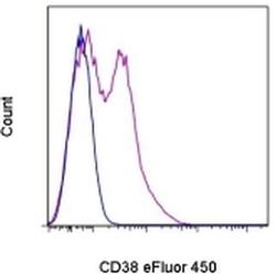
- Experimental details
- Staining of normal human peripheral blood cells with Mouse IgG1 K Isotype Control eFluor® 450 (Product # 48-4714-82) (blue histogram) or Anti-Human CD38 eFluor® 450 (purple histogram). Cells in the lymphocyte gate were used for analysis.
