 Explore
 Validate
 Learn
Learn Western blot
 Immunocytochemistry
Antibody data
- Antibody Data
- Antigen structure
- References [0]
- Comments [0]
- Validations
- Immunocytochemistry [3]
Submit
Validation data
Reference
Comment
Report error
- Product number
- SM5081 - Provider product page

- Provider
- Acris Antibodies GmbH
- Proper citation
- Acris Antibodies GmbH Cat#SM5081, RRID:AB_1005665
- Product name
- anti NFAT2 / NFATC (1-654)
- Antibody type
- Monoclonal
- Antigen
- Bacterially expressed glutathione S-transferase (GST) fusion protein containing NF-ATc1 residues 1-654.
- Reactivity
- Human, Mouse, Rat, Simian
- Host
- Mouse
- Isotype
- IgG
- Antibody clone number
- 7A6
- Vial size
- 0.1 ml
- Concentration
- This product is unpurified. The exact concentration of antibody is not quantifiable.
No comments: Submit comment
Supportive validation
- Submitted by
- Acris Antibodies GmbH (provider)
- Main image
- Experimental details
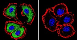
- Immunofluorescent analysis of NFATc1 using NFATc1 Monoclonal Antibody (7A6) (Cat.-No SM5081) shows staining in MCF-7 Cells. NFATc1 (green), F-Actin staining with Phalloidin (red) and nuclei with DAPI (blue) is shown. Cells were grown on chamber slides and fixed with formaldehyde prior to staining. Cells were probed without (control) or with an antibody recognizing NFATc1 (Cat.-No SM5081) at a dilution of 1:20 over night at 4 ?C, washed with PBS and incubated with a DyLight-488 conjugated secondary antibody. Images were taken at 60X magnification

- Submitted by
- Acris Antibodies GmbH (provider)
- Main image
- Experimental details
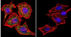
- Immunofluorescent analysis of NFATc1 using NFATc1 Monoclonal Antibody (7A6) (Cat.-No SM5081) shows staining in Hela Cells. NFATc1 (green), F-Actin staining with Phalloidin (red) and nuclei with DAPI (blue) is shown. Cells were grown on chamber slides and fixed with formaldehyde prior to staining. Cells were probed without (control) or with an antibody recognizing NFATc1 (Cat.-No SM5081) at a dilution of 1:20 over night at 4°C, washed with PBS and incubated with a DyLight-488 conjugated secondary antibody. Images were taken at 60X magnification.

- Submitted by
- Acris Antibodies GmbH (provider)
- Main image
- Experimental details
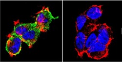
- Immunofluorescent analysis of NFATc1 using NFATc1 Monoclonal Antibody (7A6) (Cat.-No SM5081) shows staining in 293Cells. NFATc1 (green), F-Actin staining with Phalloidin (red) and nuclei with DAPI (blue) is shown. Cells were grown on chamber slides and fixed with formaldehyde prior to staining. Cells were probed without (control) or with an antibody recognizing NFATc1 (Cat.-No SM5081) at a dilution of 1:20 over night at 4°C, washed with PBS and incubated with a DyLight-488 conjugated secondary antibody. Images were taken at 60X magnification.
