 Explore
 Validate
 Learn
Learn Western blot
 Immunocytochemistry
 Immunohistochemistry
Antibody data
- Antibody Data
- Antigen structure
- References [0]
- Comments [0]
- Validations
- Immunocytochemistry [7]
Submit
Validation data
Reference
Comment
Report error
- Product number
- PA1-16729 - Provider product page

- Provider
- Invitrogen Antibodies
- Product name
- GAP43 Polyclonal Antibody
- Antibody type
- Polyclonal
- Antigen
- Synthetic peptide
- Description
- Suggested positive control: embryonic nervous tissue, brain, injured nerve. Immunogen sequence: KEDPEADQEH A
- Reactivity
- Human, Mouse, Rat, Bovine, Canine, Chicken/Avian, Drosophila, Porcine
- Host
- Rabbit
- Isotype
- IgG
- Vial size
- 100 μL
- Concentration
- 1 mg/mL
- Storage
- -20°C, Avoid Freeze/Thaw Cycles
No comments: Submit comment
Supportive validation
- Submitted by
- Invitrogen Antibodies (provider)
- Main image
- Experimental details
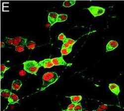
- Immunofluorescence of GAP-43 (green), a molecular marker of neurite outgrowth, demonstrates intense staining in overexpressing wild-type PS-1 (E) PC-12 cells. (Teo, et al, 2005)

- Submitted by
- Invitrogen Antibodies (provider)
- Main image

- Experimental details
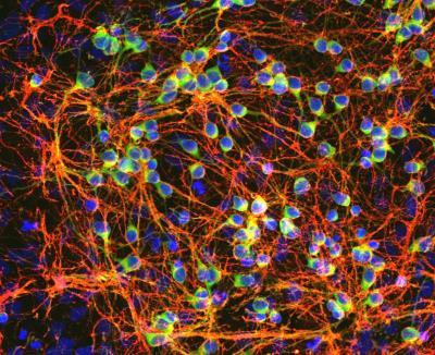
- Immunocytochemistry analysis of GAP43 in Cortical neuron-glial cell culture from E20 rat. Samples were incubated in GAP43 polyclonal antibody (Product # PA1-16729) using a dilution of 1:2000. This antibody in green, and costained with mouse mAb to vimentin, dilution 1:2,000, in red. Blue: DAPI staining of nuclear DNA. GAP43 antibody labels protein expressed in the axonal membrane of neuronal cells, while vimentin antibody stains intermediate filaments in fibroblasts and other non-neuronal cells.

- Submitted by
- Invitrogen Antibodies (provider)
- Main image

- Experimental details
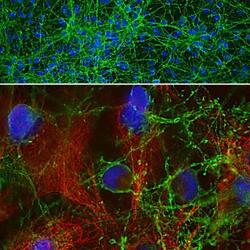
- Immunocytochemistry analysis of GAP43 in Rat E18 mixed neuron/glia cultures. Samples were incubated in GAP43 polyclonal antibody (Product # PA1-16729). GAP43 (red) and 5B10, mouse monoclonal to MAP-tau (green).
- Submitted by
- Invitrogen Antibodies (provider)
- Main image

- Experimental details
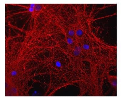
- Immunocytochemistry analysis of GAP43 in mixed neuron/glial cultures. Samples were incubated in GAP43 polyclonal antibody (Product # PA1-16729). This antibody (red), blue is DNA staining.
- Submitted by
- Invitrogen Antibodies (provider)
- Main image
- Experimental details
- Immunocytochemistry analysis of GAP43 in Cortical neuron-glial cell culture from E20 rat. Samples were incubated in GAP43 polyclonal antibody (Product # PA1-16729) using a dilution of 1:2000. This antibody in green, and costained with mouse mAb to vimentin, dilution 1:2,000, in red. Blue: DAPI staining of nuclear DNA. GAP43 antibody labels protein expressed in the axonal membrane of neuronal cells, while vimentin antibody stains intermediate filaments in fibroblasts and other non-neuronal cells.

- Submitted by
- Invitrogen Antibodies (provider)
- Main image

- Experimental details
- Immunocytochemistry analysis of GAP43 in Rat E18 mixed neuron/glia cultures. Samples were incubated in GAP43 polyclonal antibody (Product # PA1-16729). GAP43 (red) and 5B10, mouse monoclonal to MAP-tau (green).

- Submitted by
- Invitrogen Antibodies (provider)
- Main image

- Experimental details
- Immunocytochemistry analysis of GAP43 in mixed neuron/glial cultures. Samples were incubated in GAP43 polyclonal antibody (Product # PA1-16729). This antibody (red), blue is DNA staining.
