 Explore
 Validate
 Learn
Learn Western blot
 Immunocytochemistry
Antibody data
- Antibody Data
- Antigen structure
- References [1]
- Comments [0]
- Validations
- Immunocytochemistry [6]
- Immunohistochemistry [1]
- Flow cytometry [1]
- Other assay [1]
Submit
Validation data
Reference
Comment
Report error
- Product number
- MA1-19168 - Provider product page

- Provider
- Invitrogen Antibodies
- Product name
- Vimentin Monoclonal Antibody (VI-01)
- Antibody type
- Monoclonal
- Antigen
- Other
- Description
- This antibody reacts with vimentin, a 57 kDa intermediate filament intracellular protein expressed in variety of mesenchymal and mesodermal cell types. Cross-reactivity was found with smooth muscle desmin. Immunocytochemistry: Staining technique: (a) fix cells for 10 min in methanol at -20°C and for 6 min in acetone at -20°C; (b) fix cells directly in methanol for 10 min at -20°C or in acetone for 10 min at -20°C. Positive control: 3T3 murine Swiss albino fibroblast cell line, RBL rat basophilic leukemia cell line. Flow cytometry: Intracellular staining.
- Reactivity
- Human, Mouse, Porcine
- Host
- Mouse
- Isotype
- IgM
- Antibody clone number
- VI-01
- Vial size
- 100 μg
- Concentration
- 1 mg/mL
- Storage
- 4°C, do not freeze
Submitted references Bioengineered uterine tissue supports pregnancy in a rat model.
Hellström M, Moreno-Moya JM, Bandstein S, Bom E, Akouri RR, Miyazaki K, Maruyama T, Brännström M
Fertility and sterility 2016 Aug;106(2):487-496.e1
Fertility and sterility 2016 Aug;106(2):487-496.e1
No comments: Submit comment
Supportive validation
- Submitted by
- Invitrogen Antibodies (provider)
- Main image
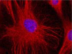
- Experimental details
- Immunofluorescent analysis of Vimentin using a monoclonal antibody (Product # MA1-19168).
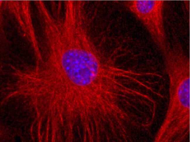
- Submitted by
- Invitrogen Antibodies (provider)
- Main image
- Experimental details
- Immunofluorescent analysis of Vimentin using a monoclonal antibody (Product # MA1-19168).
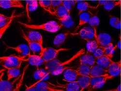
- Submitted by
- Invitrogen Antibodies (provider)
- Main image

- Experimental details
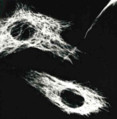
- Immunocytochemistry analysis of vimentin in methanol/acetone fixed murine 3T3 cells using mouseMonoclonal antibody VI-01 (Product # MA1-19168).
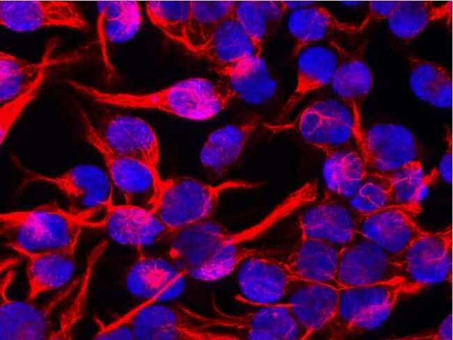
- Submitted by
- Invitrogen Antibodies (provider)
- Main image

- Experimental details
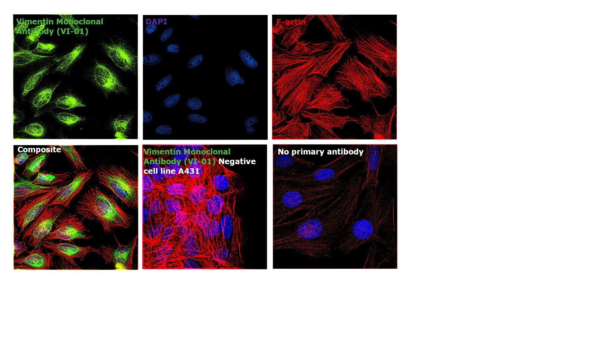
- mmunofluorescence analysis of Vimentin was performed using 70% confluent log phase HeLa cells. The cells were fixed with 4% paraformaldehyde for 10 minutes, permeabilized with 0.1% Triton™ X-100 for 10 minutes, and blocked with 1% BSA for 1 hour at room temperature. The cells were labeled with Vimentin Mouse Monoclonal Antibody (Product # MA1-19168) at 5 µg/mL in 0.1% BSA and incubated overnight at 4 degree Celsius and then labeled with Goat anti-Mouse IgG (H+L)/IgM (L) Superclonal™ Secondary Antibody, Alexa Fluor® 488 conjugate (Product # A28175) at a dilution of 1:2000 for 45 minutes at room temperature (Panel a: green). Nuclei (Panel b: blue) were stained with SlowFade® Gold Antifade Mountant with DAPI (Product # S36938). F-actin (Panel c: red) was stained with Rhodamine Phalloidin (Product # R415, 1:300). Panel d represents the merged image showing cytoplasmic, cytoskeletal and nuclear localization. Panel e represents negative control, A-431 cells. Panel f represents control cells with no primary antibody to assess background. The images were captured at 60X magnification.
- Submitted by
- Invitrogen Antibodies (provider)
- Main image

- Experimental details
- Immunocytochemistry analysis of vimentin in methanol/acetone fixed murine 3T3 cells using mouseMonoclonal antibody VI-01 (Product # MA1-19168).

- Submitted by
- Invitrogen Antibodies (provider)
- Main image

- Experimental details
- mmunofluorescence analysis of Vimentin was performed using 70% confluent log phase HeLa cells. The cells were fixed with 4% paraformaldehyde for 10 minutes, permeabilized with 0.1% Triton™ X-100 for 10 minutes, and blocked with 1% BSA for 1 hour at room temperature. The cells were labeled with Vimentin Mouse Monoclonal Antibody (Product # MA1-19168) at 5 µg/mL in 0.1% BSA and incubated overnight at 4 degree Celsius and then labeled with Goat anti-Mouse IgG (H+L)/IgM (L) Superclonal™ Secondary Antibody, Alexa Fluor® 488 conjugate (Product # A28175) at a dilution of 1:2000 for 45 minutes at room temperature (Panel a: green). Nuclei (Panel b: blue) were stained with SlowFade® Gold Antifade Mountant with DAPI (Product # S36938). F-actin (Panel c: red) was stained with Rhodamine Phalloidin (Product # R415, 1:300). Panel d represents the merged image showing cytoplasmic, cytoskeletal and nuclear localization. Panel e represents negative control, A-431 cells. Panel f represents control cells with no primary antibody to assess background. The images were captured at 60X magnification.

Supportive validation
- Submitted by
- Invitrogen Antibodies (provider)
- Main image

- Experimental details
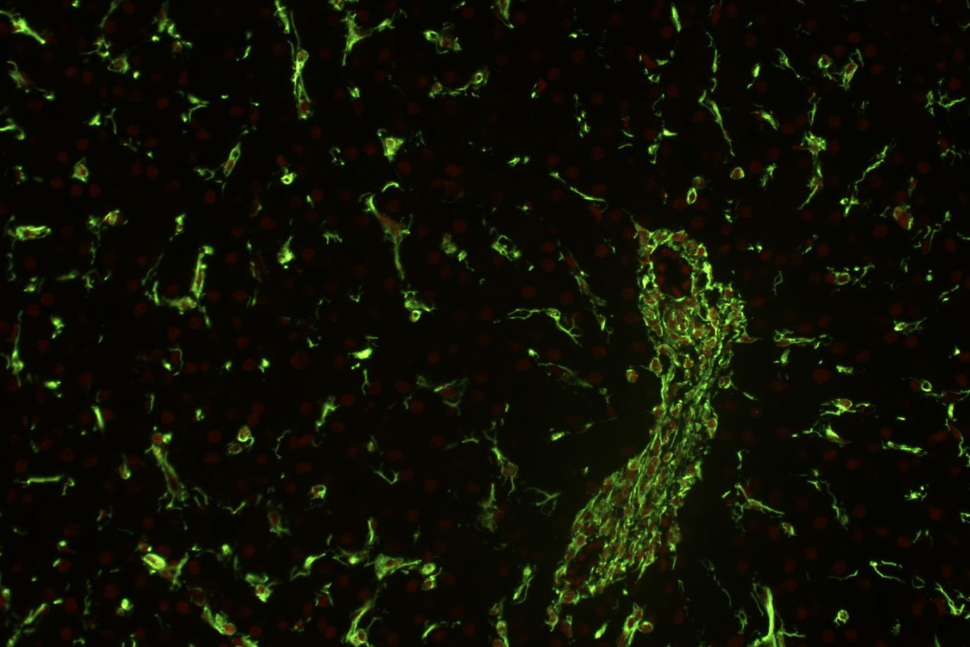
- Immunohistochemistry (Paraffin) analysis of Vimentin using Vimentin Monoclonal Antibody (VI-01) (Product # MA1-19168) in human liver at a dilution of 1:400, detected with GAM IgM-Alexa Fluor™488 (diluted 1:200; green), cell nuclei stained with PI (1 µg/mL; orange).
Supportive validation
- Submitted by
- Invitrogen Antibodies (provider)
- Main image
- Experimental details
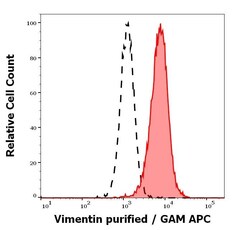
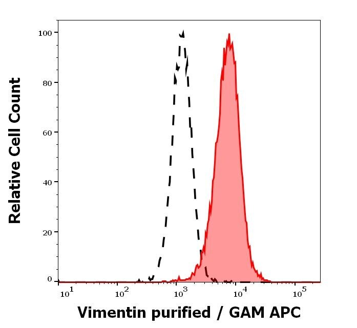
- Flow Cytometry analysis of Vimentin using Vimentin Monoclonal Antibody (VI-01) (Product # MA1-19168). Separation of HeLa cells stained using anti-Vimentin (VI-01) purified antibody (concentration in sample 5 μg/mL, GAM APC, red-filled) from HeLa cells unstained by primary antibody (GAM APC, black-dashed) in flow cytometry analysis (intracellular staining of methanol permeabilisated cells).
Supportive validation
- Submitted by
- Invitrogen Antibodies (provider)
- Main image
- Experimental details
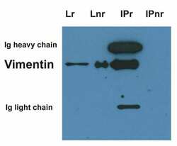
- Immunoprecipitation of Vimentin using a monoclonal antibody (Product # MA1-19168).
