 Explore
 Validate
 Learn
Learn Western blot
Antibody data
- Antibody Data
- Antigen structure
- References [0]
- Comments [0]
- Validations
- Western blot [4]
Submit
Validation data
Reference
Comment
Report error
- Product number
- PA5-29075 - Provider product page

- Provider
- Invitrogen Antibodies
- Product name
- PAK3 Polyclonal Antibody
- Antibody type
- Polyclonal
- Antigen
- Recombinant full-length protein
- Description
- Recommended positive controls: NT2D1, IMR32, U87-MG, MCF-7, NIH-3T3, JC, mouse brain, rat brain. Predicted reactivity: Mouse (100%), Rat (100%), Xenopus laevis (99%), Chimpanzee (100%), Bovine (100%). Store product as a concentrated solution. Centrifuge briefly prior to opening the vial.
- Reactivity
- Human, Mouse, Rat
- Host
- Rabbit
- Isotype
- IgG
- Vial size
- 100 μL
- Concentration
- 0.74 mg/mL
- Storage
- Store at 4°C short term. For long term storage, store at -20°C, avoiding freeze/thaw cycles.
No comments: Submit comment
Supportive validation
- Submitted by
- Invitrogen Antibodies (provider)
- Main image
- Experimental details
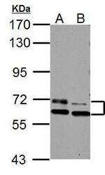
- Western Blot using PAK3 Polyclonal Antibody (Product # PA5-29075). Sample (30 µg of whole cell lysate). Lane A: NIH-3T3. Lane B: BCL-1 . 7.5% SDS PAGE. PAK3 Polyclonal Antibody (Product # PA5-29075) diluted at 1:1,000.

- Submitted by
- Invitrogen Antibodies (provider)
- Main image
- Experimental details
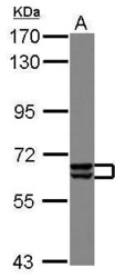
- Western Blot analysis of PAK3 was performed by separating 30 µg of IMR32 lysates by 7.5% SDS PAGE. Proteins were transferred to a membrane and probed with a PAK3 Polyclonal Antibody (Product # PA5-29075) at a dilution of 1:1000.

- Submitted by
- Invitrogen Antibodies (provider)
- Main image
- Experimental details
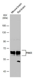
- Western Blot analysis of PAK3 was performed by separating 50 µg of Various tissue extracts by 7.5% SDS-PAGE. Proteins were transferred to a membrane and probed with a PAK3 Polyclonal Antibody (Product # PA5-29075) at a dilution of 1:1000. The HRP-conjugated anti-rabbit IgG antibody was used to detect the primary antibody.

- Submitted by
- Invitrogen Antibodies (provider)
- Main image
- Experimental details
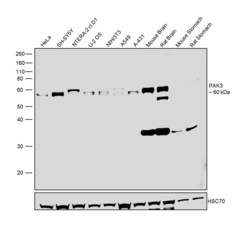
- Western blot was performed using Anti-PAK3 Polyclonal Antibody (Product # PA5-29075) and a 60 kDa band corresponding to PAK3 was observed across the cell lines and tissues tested except Mouse and Rat Stomach. Whole cell extracts (30 µg lysate) of HeLa (Lane 1), SH-SY5Y (Lane 2), NTERA-2 cl.D1 (Lane 3), U-2 OS (Lane 4), NIH/3T3 (Lane 5), A549 (Lane 6), A-431 (Lane 7), tissue extracts of Mouse Brain (Lane 8), Rat Brain (Lane 9), Mouse Stomach (Lane 10) and Rat Stomach (Lane 11) were electrophoresed using NuPAGE™ 10% Bis-Tris Protein Gel (Product # NP0302BOX). Resolved proteins were then transferred onto a nitrocellulose membrane (Product # IB23001) by iBlot® 2 Dry Blotting System (Product # IB21001). The blot was probed with the primary antibody (1:500 dilution) and detected by chemiluminescence with Goat anti-Rabbit IgG (Heavy Chain), Superclonal™ Recombinant Secondary Antibody, HRP (Product # A27036, 1:4000 dilution) using the iBright FL 1000 (Product # A32752). Chemiluminescent detection was performed using SuperSignal™ West Dura Extended Duration Substrate (Product # 34076).
