 Explore
 Validate
 Learn
Learn Western blot
 ELISA
ELISA Immunocytochemistry
Antibody data
- Antibody Data
- Antigen structure
- References [0]
- Comments [0]
- Validations
- Immunocytochemistry [2]
Submit
Validation data
Reference
Comment
Report error
- Product number
- MA1-19178 - Provider product page

- Provider
- Invitrogen Antibodies
- Product name
- AFP Monoclonal Antibody (AFP-01)
- Antibody type
- Monoclonal
- Antigen
- Purifed from natural sources
- Description
- This antibody reacts with human alpha-Fetoprotein (AFP), a 70 kDa oncofetal antigen. AFP is a major fetal plasma protein, but is not present in healthy adult tissues. Elevated AFP concentrations in adult plasma may be an early marker of hepatocellular carcinoma or teratoblastoma, while high concentrations in amniotic fluid may indicate severe congenital defects of a fetus. This antibody has successfully been paired as the coating antibody in a sandwich ELISA with detection antibody MA1-19178 (clone AFP-01). Immunoprecipitation: Interaction of the antibody AFP-01 with AFP is dependent on the presence of calcium ions (strongly inhibited by chelating agents). Such characteristics of the antibody can be exploited for immunoaffinity purification of APF under mild elution conditions; Western Blot: non-reducing conditions.
- Reactivity
- Human
- Host
- Mouse
- Isotype
- IgG
- Antibody clone number
- AFP-01
- Vial size
- 100 μg
- Concentration
- 1 mg/mL
- Storage
- 4°C, do not freeze
No comments: Submit comment
Supportive validation
- Submitted by
- Invitrogen Antibodies (provider)
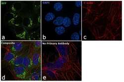
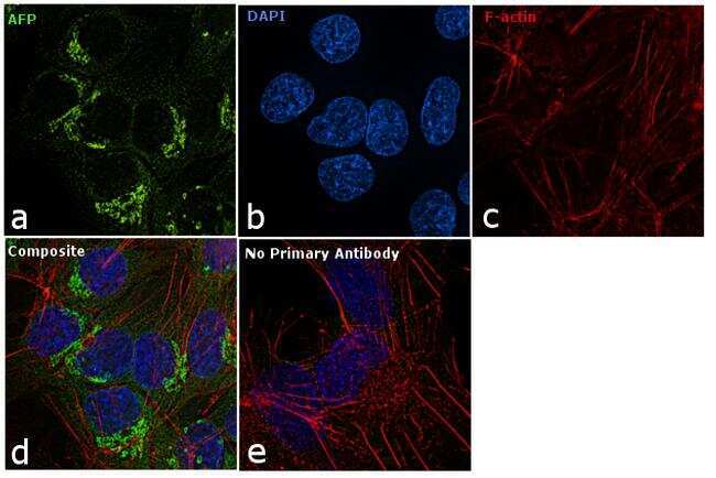
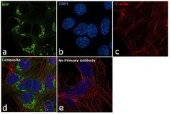
- Main image
- Experimental details
- Immunofluorescence analysis of AFP was performed using 70% confluent log phase Hep G2 cells. The cells were fixed with 4% paraformaldehyde for 10 minutes, permeabilized with 0.1% Triton™ X-100 for 15 minutes, and blocked with 1% BSA for 1 hour at room temperature. The cells were labeled with AFP Mouse Monoclonal Antibody (Product # MA1-19178) at 5 µg/mL in 0.1% BSA, incubated at 4 degree Celsius overnight and then labeled with Goat anti-Mouse IgG (H+L) Superclonal™ Secondary Antibody, Alexa Fluor® 488 conjugate (Product # A28175) at a dilution of 1:2000 for 45 minutes at room temperature (Panel a: green). Nuclei (Panel b: blue) were stained with ProLong™ Diamond Antifade Mountant with DAPI (Product # P36962). F-actin (Panel c: red) was stained with Rhodamine Phalloidin (Product # R415, 1:300). Panel d represents the merged image showing cytoplasmic and Golgi localization. Panel e represents control cells with no primary antibody to assess background. The images were captured at 60X magnification.
- Submitted by
- Invitrogen Antibodies (provider)
- Main image
- Experimental details
- Immunofluorescence analysis of AFP was performed using 70% confluent log phase Hep G2 cells. The cells were fixed with 4% paraformaldehyde for 10 minutes, permeabilized with 0.1% Triton™ X-100 for 15 minutes, and blocked with 1% BSA for 1 hour at room temperature. The cells were labeled with AFP Mouse Monoclonal Antibody (Product # MA1-19178) at 5 µg/mL in 0.1% BSA, incubated at 4 degree Celsius overnight and then labeled with Goat anti-Mouse IgG (H+L) Superclonal™ Secondary Antibody, Alexa Fluor® 488 conjugate (Product # A28175) at a dilution of 1:2000 for 45 minutes at room temperature (Panel a: green). Nuclei (Panel b: blue) were stained with ProLong™ Diamond Antifade Mountant with DAPI (Product # P36962). F-actin (Panel c: red) was stained with Rhodamine Phalloidin (Product # R415, 1:300). Panel d represents the merged image showing cytoplasmic and Golgi localization. Panel e represents control cells with no primary antibody to assess background. The images were captured at 60X magnification.
