 Explore
 Validate
 Learn
Learn Western blot
 ELISA
ELISA Immunocytochemistry
Antibody data
- Antibody Data
- Antigen structure
- References [0]
- Comments [0]
- Validations
- Immunocytochemistry [2]
Submit
Validation data
Reference
Comment
Report error
- Product number
- MA1-35436 - Provider product page

- Provider
- Invitrogen Antibodies
- Product name
- AFP Monoclonal Antibody (C2)
- Antibody type
- Monoclonal
- Antigen
- Other
- Description
- C2 has been characterized in the ISOBM TD-2 workshop and assigned by K. Nustad to group E of a cluster of 6 major epitopes of human alpha fetoprotein. Human alpha fetoprotein is an oncofetal protein of 70 kDa. It is expressed in fetal liver and is normally absent in health adult tissues. It is positive on all yolk sac tumors, on some other germ cell tumors and on hepatocellular carcinomas.
- Reactivity
- Human, Mouse
- Host
- Mouse
- Isotype
- IgG
- Antibody clone number
- C2
- Vial size
- 100 μg
- Concentration
- 100 μg/mL
- Storage
- 4°C
No comments: Submit comment
Supportive validation
- Submitted by
- Invitrogen Antibodies (provider)
- Main image
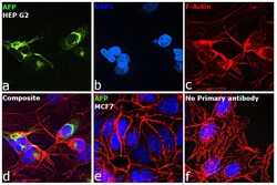
- Experimental details
- Immunofluorescence analysis of AFP was performed using 70% confluent log phase Hep G2 cells and MCF7 cells. The cells were fixed with 4% paraformaldehyde for 10 minutes, permeabilized with 0.1% Triton™ X-100 for 15 minutes, and blocked with 2% BSA for 1 hour at room temperature. The cells were labeled with AFP Monoclonal Antibody (C2) (Product # MA1-35436) at 1:100 in 0.1% BSA, incubated at 4 degree celsius overnight and then labeled with Donkey anti-Mouse IgG (H+L) Highly Cross-Adsorbed Secondary Antibody, Alexa Fluor™ Plus 488 (Product # A32766, 1:2,000), for 45 minutes at room temperature (Panel a: Green). Nuclei (Panel b: Blue) were stained with ProLong™ Diamond Antifade Mountant with DAPI (Product # P36962). F-actin (Panel c: Red) was stained with Rhodamine Phalloidin (Product # R415, 1:300). Panel d represents the merged image showing cytoplasmic and golgi staining of AFP in Hep G2 cells. Panel e represents MCF7 with no signal. Panel f represents control cells with no primary antibody to assess background. The images were captured at 60X magnification.

- Submitted by
- Invitrogen Antibodies (provider)
- Main image
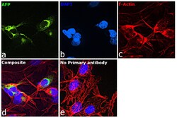
- Experimental details
- Immunofluorescence analysis of AFP was performed using 70% confluent log phase Hep G2 cells. The cells were fixed with 4% paraformaldehyde for 10 minutes, permeabilized with 0.1% Triton™ X-100 for 15 minutes, and blocked with 2% BSA for 1 hour at room temperature. The cells were labeled with AFP Monoclonal Antibody (C2) (Product # MA1-35436) at 1:100 in 0.1% BSA, incubated at 4 degree celsius overnight and then labeled with Donkey anti-Mouse IgG (H+L) Highly Cross-Adsorbed Secondary Antibody, Alexa Fluor™ Plus 488 (Product # A32766, 1:2,000), for 45 minutes at room temperature (Panel a: Green). Nuclei (Panel b:Blue) were stained with ProLong™ Diamond Antifade Mountant with DAPI (Product # P36962). F-actin (Panel c: Red) was stained with Rhodamine Phalloidin (Product # R415, 1:300). Panel d represents the merged image showing cytoplasmic and golgi staining of AFP in Hep G2 cells. Panel e represents control cells with no primary antibody to assess background. The images were captured at 60X magnification.
