 Explore
 Validate
 Learn
Learn Western blot
 Immunocytochemistry
Antibody data
- Antibody Data
- Antigen structure
- References [0]
- Comments [0]
- Validations
- Immunocytochemistry [7]
- Immunohistochemistry [5]
Submit
Validation data
Reference
Comment
Report error
- Product number
- PA5-29557 - Provider product page

- Provider
- Invitrogen Antibodies
- Product name
- ApoA1 Polyclonal Antibody
- Antibody type
- Polyclonal
- Antigen
- Recombinant full-length protein
- Description
- Recommended positive controls: human plasma, mouse plasma, rat plasma. Predicted reactivity: Pig (80%). Store product as a concentrated solution. Centrifuge briefly prior to opening the vial.
- Reactivity
- Human, Mouse, Rat
- Host
- Rabbit
- Isotype
- IgG
- Vial size
- 100 μL
- Concentration
- 1.43 mg/mL
- Storage
- Store at 4°C short term. For long term storage, store at -20°C, avoiding freeze/thaw cycles.
No comments: Submit comment
Supportive validation
- Submitted by
- Invitrogen Antibodies (provider)
- Main image

- Experimental details
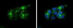
- ApoA1 Polyclonal Antibody detects Apolipoprotein A1 protein at Golgi apparatus by immunofluorescent analysis. Sample: HepG2 cells were fixed in 4% paraformaldehyde at RT for 15 min. Green: Apolipoprotein A1 stained by ApoA1 Polyclonal Antibody (Product # PA5-29557) diluted at 1:500. Red: alpha Tubulin, a cytoskeleton marker, stained by alpha Tubulin Polyclonal Antibody [GT114] (Product # MA5-31466) diluted at 1:1,000. Blue: Fluoroshield with DAPI .

- Submitted by
- Invitrogen Antibodies (provider)
- Main image
- Experimental details
- Immunocytochemistry-Immunofluorescence analysis of ApoA1 was performed in HepG2 cells fixed in 4% paraformaldehyde at RT for 15 min. Green: ApoA1 Polyclonal Antibody (Product # PA5-29557) diluted at 1:500. Blue: Hoechst 33342 staining.

- Submitted by
- Invitrogen Antibodies (provider)
- Main image

- Experimental details
- ApoA1 Polyclonal Antibody detects Apolipoprotein A1 protein at Golgi apparatus by immunofluorescent analysis. Sample: HepG2 cells were fixed in 4% paraformaldehyde at RT for 15 min. Green: Apolipoprotein A1 stained by ApoA1 Polyclonal Antibody (Product # PA5-29557) diluted at 1:500. Red: alpha Tubulin, a cytoskeleton marker, stained by alpha Tubulin Polyclonal Antibody [GT114] (Product # MA5-31466) diluted at 1:1,000. Blue: Fluoroshield with DAPI .

- Submitted by
- Invitrogen Antibodies (provider)
- Main image
- Experimental details
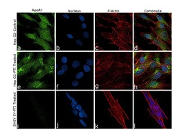
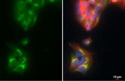
- Immunofluorescence analysis using Anti-ApoA1 Polyclonal Antibody (Product # PA5-29557), shows higher expression of ApoA1 in Hep G2 cell line as compared to SH-SY5Y. The cells were fixed with 4% paraformaldehyde for 10 minutes, permeabilized with 0.1% Triton™ X-100 for 15 minutes, and blocked with 2% BSA for 45 minutes at room temperature. The cells were labeled with ApoA1 Polyclonal Antibody (Product # PA5-29557) at 5 µg in 0.1% BSA, incubated at 4 degree celsius overnight and then labeled with Goat anti-Rabbit IgG (H+L) Superclonal™ Recombinant Secondary Antibody, Alexa Fluor® 488 conjugate (Product # A27034), (1:2000 dilution), for 45 minutes at room temperature (Panel a: Blue). Nuclei (Panel b: Blue) were stained with ProLong™ Diamond Antifade Mountant with DAPI (Product # P36962). F-actin (Panel c: Red) was stained with Rhodamine Phalloidin (Product # R415, 1:300 dilution). Panel d represents the merged image of untreated HepG2 cells showing staining for APOA1 that is enhanced upon PTI treatment (Panel h). Panel l represents SH-SY 5Y cells showing no staining for APOA1 upon PTI treatment. The images were captured at 60X magnification. APOA1 is secretory protein showing cytoplasmic and Golgi apparatus expression as seen in panel d and h respectively upon PTI treatment.

- Submitted by
- Invitrogen Antibodies (provider)
- Main image
- Experimental details
- ApoA1 Polyclonal Antibody detects Apolipoprotein A1 protein at Golgi apparatus by immunofluorescent analysis. Sample: HepG2 cells were fixed in 4% paraformaldehyde at RT for 15 min. Green: Apolipoprotein A1 stained by ApoA1 Polyclonal Antibody (Product # PA5-29557) diluted at 1:500. Red: alpha Tubulin, a cytoskeleton marker, stained by alpha Tubulin Polyclonal Antibody [GT114] (Product # MA5-31466) diluted at 1:1,000. Blue: Fluoroshield with DAPI .

- Submitted by
- Invitrogen Antibodies (provider)
- Main image

- Experimental details
- Immunocytochemistry-Immunofluorescence analysis of ApoA1 was performed in HepG2 cells fixed in 4% paraformaldehyde at RT for 15 min. Green: ApoA1 Polyclonal Antibody (Product # PA5-29557) diluted at 1:500. Blue: Hoechst 33342 staining.

- Submitted by
- Invitrogen Antibodies (provider)
- Main image

- Experimental details
- Immunofluorescence analysis using Anti-ApoA1 Polyclonal Antibody (Product # PA5-29557), shows higher expression of ApoA1 in Hep G2 cell line as compared to SH-SY5Y. The cells were fixed with 4% paraformaldehyde for 10 minutes, permeabilized with 0.1% Triton™ X-100 for 15 minutes, and blocked with 2% BSA for 45 minutes at room temperature. The cells were labeled with ApoA1 Polyclonal Antibody (Product # PA5-29557) at 5 µg in 0.1% BSA, incubated at 4 degree celsius overnight and then labeled with Goat anti-Rabbit IgG (Heavy Chain) Superclonal™ Recombinant Secondary Antibody, Alexa Fluor® 488 conjugate (Product # A27034), (1:2000 dilution), for 45 minutes at room temperature (Panel a: Blue). Nuclei (Panel b: Blue) were stained with ProLong™ Diamond Antifade Mountant with DAPI (Product # P36962). F-actin (Panel c: Red) was stained with Rhodamine Phalloidin (Product # R415, 1:300 dilution). Panel d represents the merged image of untreated HepG2 cells showing staining for APOA1 that is enhanced upon PTI treatment (Panel h). Panel l represents SH-SY 5Y cells showing no staining for APOA1 upon PTI treatment. The images were captured at 60X magnification. APOA1 is secretory protein showing cytoplasmic and Golgi apparatus expression as seen in panel d and h respectively upon PTI treatment.

Supportive validation
- Submitted by
- Invitrogen Antibodies (provider)
- Main image
- Experimental details
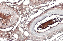
- ApoA1 Polyclonal Antibody detects secreted Apolipoprotein A1 protein by immunohistochemical analysis. Sample: Paraffin-embedded human esophagus. Apolipoprotein A1 stained by ApoA1 Polyclonal Antibody (Product # PA5-29557) diluted at 1:500. Antigen Retrieval: Citrate buffer, pH 6.0, 15 min.

- Submitted by
- Invitrogen Antibodies (provider)
- Main image
- Experimental details
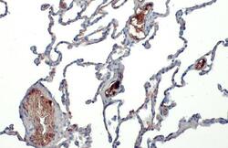
- ApoA1 Polyclonal Antibody detects secreted Apolipoprotein A1 protein by immunohistochemical analysis. Sample: Paraffin-embedded human lung. Apolipoprotein A1 stained by ApoA1 Polyclonal Antibody (Product # PA5-29557) diluted at 1:500. Antigen Retrieval: Citrate buffer, pH 6.0, 15 min.

- Submitted by
- Invitrogen Antibodies (provider)
- Main image
- Experimental details
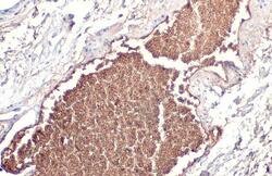
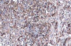
- ApoA1 Polyclonal Antibody detects secreted Apolipoprotein A1 protein by immunohistochemical analysis. Sample: Paraffin-embedded human ovarian cancer. Apolipoprotein A1 stained by ApoA1 Polyclonal Antibody (Product # PA5-29557) diluted at 1:1,000. Antigen Retrieval: Citrate buffer, pH 6.0, 15 min.

- Submitted by
- Invitrogen Antibodies (provider)
- Main image
- Experimental details
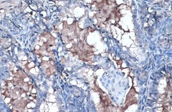
- ApoA1 Polyclonal Antibody detects secreted Apolipoprotein A1 protein by immunohistochemical analysis. Sample: Paraffin-embedded human colon. Apolipoprotein A1 stained by ApoA1 Polyclonal Antibody (Product # PA5-29557) diluted at 1:500. Antigen Retrieval: Citrate buffer, pH 6.0, 15 min.

- Submitted by
- Invitrogen Antibodies (provider)
- Main image
- Experimental details
- Immunohistochemistry (Paraffin) analysis of ApoA1 was performed in paraffin-embedded human ovarian cancer tissue using ApoA1 Polyclonal Antibody (Product # PA5-29557) at a dilution of 1:500. Antigen Retrieval: Citrate buffer, pH 6.0, 15 min.
