 Explore
 Validate
 Learn
Learn Western blot
 Immunocytochemistry
Antibody data
- Antibody Data
- Antigen structure
- References [0]
- Comments [0]
- Validations
- Immunocytochemistry [3]
- Flow cytometry [3]
Submit
Validation data
Reference
Comment
Report error
- Product number
- MA1-024 - Provider product page

- Provider
- Invitrogen Antibodies
- Product name
- Anti-TRA-1-81
- Antibody type
- Monoclonal
- Antigen
- human embryonal carcinoma cell line 2102Ep
- Reactivity
- Human
- Host
- Mouse
- Isotype
- IgM
- Antibody clone number
- tra-1-81
- Vial size
- 100 ug
- Concentration
- 1 mg/ml
- Storage
- -20° C, Avoid Freeze/Thaw Cycles
No comments: Submit comment
Supportive validation
- Submitted by
- Invitrogen Antibodies (provider)
- Main image
- Experimental details
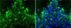
- Immunofluorescent analysis of TRA-1-81 using anti-TRA-1-81 monoclonal antibody (Product # MA1-024) shows staining on the cell surface of human H9 human embryonic stem cells, indicating pluripotency. TRA-1-81 staining (green) and an overlay image of TRA-1-81 with DAPI (blue) is shown. H9 human embryonic stem cells were grown on matrigel coated chamber slides and fixed with formaldehyde prior to staining. Cells were probed with a Monoclonal antibody recognizing TRA-1-81 (Product # MA1-024) at a dilution of 1:20 overnight at 4 °C, washed with PBS and incubated with a FITC-conjugated secondary antibody at a dilution of 1:100 for 60 minutes at room temperature. Images were taken at 20X magnification.

- Submitted by
- Invitrogen Antibodies (provider)
- Main image
- Experimental details
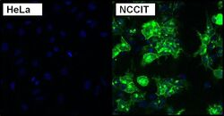
- Immunofluorescent analysis of TRA-1-81 using anti-TRA-1-81 monoclonal antibody (Product # MA1-024) shows expression in human teratocarcinoma NCCIT cells (shown in green) but not in negative control HeLa cells. Formalin fixed cells were permeabilized with 0.1% Triton X-100 in TBS for 10 minutes at room temperature. Cells were blocked with 1% Blocker BSA (Product # 37525) for 15 minutes at room temperature. Cells were probed with a mouse monoclonal antibody recognizing TRA-1-81 (Product # MA1-024), at a dilution of 1:50 for at least 1 hour at room temperature. Cells were washed with PBS and incubated with goat-anti-mouse IgM secondary antibody at a dilution of 1:400 for 30 minutes at room temperature. Nuclei (blue) were stained with Hoechst 33342 dye (Product # 62249). Images were taken on a Thermo Scientific ArrayScan at 20X magnification.

- Submitted by
- Invitrogen Antibodies (provider)
- Main image
- Experimental details
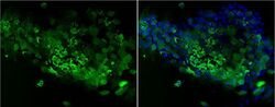
- Immunofluorescent analysis of TRA-1-81 using anti-TRA-1-81 monoclonal antibody (Product # MA1-024) shows staining on the cell surface of human HEL 11.4 iPS cells, indicating pluripotency. TRA-1-81 staining (green) and an overlay image of TRA-1-81 with DAPI (blue) is shown. HEL 11.4 iPS cells were grown on matrigel coated chamber slides and fixed with formaldehyde prior to staining. Cells were probed with a Monoclonal antibody recognizing TRA-1-81 (Product # MA1-024) at a dilution of 1:20 overnight at 4 °C, washed with PBS and incubated with a FITC-conjugated secondary antibody at a dilution of 1:100 for 60 minutes at room temperature. Images were taken at 20X magnification.

Supportive validation
- Submitted by
- Invitrogen Antibodies (provider)
- Main image
- Experimental details
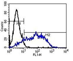
- Flow cytometry analysis of TRA-1-81 was performed on NCCIT cells. NCCIT cells were harvested, fixed and washed with PBS. Cells were incubated with 0.4 µg of TRA-1-60 monoclonal antibody (Product # MA1-024), followed by incubation with FITC-conjugated goat-anti-mouse IgM secondary antibody (Product # 31992), indicated by the shifted peak (blue line). To control for non-specific binding, cells were also incubated with secondary antibody alone (black line). Incubations were performed for 30 mins in the dark and 10,000 cells were analyzed for each sample.

- Submitted by
- Invitrogen Antibodies (provider)
- Main image
- Experimental details
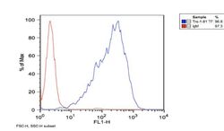
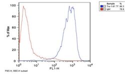
- Flow cytometry analysis of TRA-1-81 using anti-TRA-1-81 monoclonal antibody (Product # MA1-024) shows positive staining of H9 human embryonic stem cells (blue line). H9 human embryonic stem cells were harvested, fixed and washed with PBS. Cells were incubated with anti-TRA-1-81 monoclonal antibody (Product # MA1-024) or control at a 1:100 dilution for 1 hour on ice, followed by 30 min incubation with FITC-conjugated secondary antibody. 100,000 cells were stained for each sample.

- Submitted by
- Invitrogen Antibodies (provider)
- Main image
- Experimental details
- Flow cytometry analysis of TRA-1-81 using anti-TRA-1-81 monoclonal antibody (Product # MA1-024) shows positive staining of HEL 11.4 iPS cells (blue line). HEL 11.4 iPS cells were harvested, fixed and washed with PBS. Cells were incubated with anti-TRA-1-81 monoclonal antibody (Product # MA1-024) or control at a 1:100 dilution for 1 hour on ice, followed by 30 min incubation with FITC-conjugated secondary antibody. 100,000 cells were stained for each sample.
