 Explore
 Validate
 Learn
Learn Western blot
Antibody data
- Antibody Data
- Antigen structure
- References [0]
- Comments [0]
- Validations
- Western blot [2]
- Immunocytochemistry [7]
- Chromatin Immunoprecipitation [2]
Submit
Validation data
Reference
Comment
Report error
- Product number
- MA5-15185 - Provider product page

- Provider
- Invitrogen Antibodies
- Product name
- Phospho-ATM (Ser1981) Monoclonal Antibody (C.70.6)
- Antibody type
- Monoclonal
- Antigen
- Synthetic peptide
- Description
- It is not recommended to aliquot this antibody. This antibody was orginally validated as part of a Thermo Scientific Cellomics High Content Screening Kit. The antibody sold separately may have slightly different performance and may need to be further optimized for the best results.
- Reactivity
- Human
- Host
- Mouse
- Isotype
- IgG
- Antibody clone number
- C.70.6
- Vial size
- 100 μL
- Concentration
- 2028 μg/mL
- Storage
- -20°C
No comments: Submit comment
Supportive validation
- Submitted by
- Invitrogen Antibodies (provider)
- Main image
- Experimental details
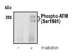
- Western blot analysis of Phospho-ATM (Ser1981) using a monoclonal antibody (Product # MA5-15185).

- Submitted by
- Invitrogen Antibodies (provider)
- Main image
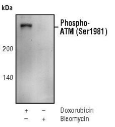
- Experimental details
- Western blot analysis of Phospho-ATM pSer1981 in extracts from M059J cells, treated with doxorubicin or treated with bleomycin, using Phospho-ATM pSer1981 monoclonal antibody (Product # MA5-15185).

Supportive validation
- Submitted by
- Invitrogen Antibodies (provider)
- Main image
- Experimental details
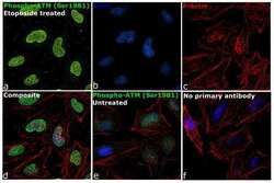
- Immunofluorescence analysis of Phospho-ATM (Ser1981) was performed using 70% confluent log phase HeLa cells treated with 10 uM Etoposide for 2 hours. The cells were fixed with 4% paraformaldehyde for 10 minutes, permeabilized with 0.1% Triton™ X-100 for 10 minutes, and blocked with 1% BSA for 1 hour at room temperature. The cells were labeled with Phospho-ATM (Ser1981) Monoclonal Antibody (C.70.6) (Product # MA5-15185) at 1:100 dilution in 0.1% BSA, incubated overnight at 4 degree Celsius and then labeled with Goat anti-Mouse IgG (H+L) Superclonal™ Secondary Antibody, Alexa Fluor® 488 conjugate (Product # A28175) at a dilution of 1:2000 for 45 minutes at room temperature (Panel a: green). Nuclei (Panel b: blue) were stained with ProLong™ Diamond Antifade Mountant with DAPI (Product # P36962). F-actin (Panel c: red) was stained with Rhodamine Phalloidin (Product # R415, 1:300). Panel d represents the merged image showing nuclear localization upon Etoposide treatment. Panel e shows untreated cells without any staining. Panel f represents control cells with no primary antibody to assess background. The images were captured at 60X magnification.

- Submitted by
- Invitrogen Antibodies (provider)
- Main image
- Experimental details
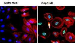
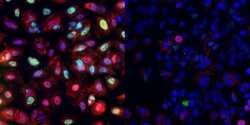
- Immunofluorescent analysis of Phospho-ATM (green) in HeLa cells either left untreated (left panel) or treated with 50uM etoposide for 3 hours (right panel). Formalin fixed cells were permeabilized with 0.1% Triton X-100 in TBS for 10 minutes at room temperature and blocked with 1% Blocker BSA (Product # 37525) for 15 minutes at room temperature. Cells were probed with a Phospho-ATM monoclonal antibody (Product # MA5-15185) at a dilution of 1:100 for at least 1 hour at room temperature, washed with PBS, and incubated with DyLight 488 goat anti-mouse IgG secondary antibody (Product # 35502) at a dilution of 1:400 for 30 minutes at room temperature. F-Actin (red) was stained with DyLight 554 phalloidin (Product # 21834) and nuclei (blue) were stained with Hoechst 33342 dye (Product # 62249). Images were taken on a Thermo Scientific ArrayScan or a ToxInsight Instrument at 20X magnification.

- Submitted by
- Invitrogen Antibodies (provider)
- Main image

- Experimental details
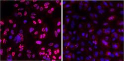
- Immunofluorescence staining of Phospho-ATM in A549 cells. Cells were treated with vehicle (0.1% DMSO in media) or with 50 µM etoposide for 3 hours

- Submitted by
- Invitrogen Antibodies (provider)
- Main image
- Experimental details
- Activation of ATM and p53: ATM kinase is auto-phosphorylated in response to DNA damage and is involved in DNA damage induced activation of p53 and cell cycle arrest. This is a multiplex kit, enabling the user to simultaneously measure both phospho-ATM and p53. A549 cells were treated with vehicle (DMSO) or 50 µM etoposide for 24 hrs. After staining cells with Cellomics phospho-ATM/p53 duplex activation kit, images were acquired and analyzed. Z-factor for phospho-ATM = 0.46 ± 0.11 and %COV = 11 ± 3. Z-factor for p53 = 0.44 ± 0.03 and %COV = 13 ± 1. EC50 of etoposide treatment for ATM activation is 19 µM and EC50 for p53 activation is 36 µM. Etoposide is a topoisomerase II inhibitor resulting in DNA strand break

- Submitted by
- Invitrogen Antibodies (provider)
- Main image

- Experimental details
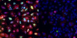
- Immunofluorescent analysis of Phospho-ATM (green) in HeLa cells either left untreated (left panel) or treated with 50uM etoposide for 3 hours (right panel). Formalin fixed cells were permeabilized with 0.1% Triton X-100 in TBS for 10 minutes at room temperature and blocked with 1% Blocker BSA (Product # 37525) for 15 minutes at room temperature. Cells were probed with a Phospho-ATM monoclonal antibody (Product # MA5-15185) at a dilution of 1:100 for at least 1 hour at room temperature, washed with PBS, and incubated with DyLight 488 goat anti-mouse IgG secondary antibody (Product # 35502) at a dilution of 1:400 for 30 minutes at room temperature. F-Actin (red) was stained with DyLight 554 phalloidin (Product # 21834) and nuclei (blue) were stained with Hoechst 33342 dye (Product # 62249). Images were taken on a Thermo Scientific ArrayScan or a ToxInsight Instrument at 20X magnification.

- Submitted by
- Invitrogen Antibodies (provider)
- Main image
- Experimental details
- Immunofluorescence staining of Phospho-ATM in A549 cells. Cells were treated with vehicle (0.1% DMSO in media) or with 50 µM etoposide for 3 hours

- Submitted by
- Invitrogen Antibodies (provider)
- Main image
- Experimental details
- Activation of ATM and p53: ATM kinase is auto-phosphorylated in response to DNA damage and is involved in DNA damage induced activation of p53 and cell cycle arrest. This is a multiplex kit, enabling the user to simultaneously measure both phospho-ATM and p53. A549 cells were treated with vehicle (DMSO) or 50 µM etoposide for 24 hrs. After staining cells with Cellomics phospho-ATM/p53 duplex activation kit, images were acquired and analyzed. Z-factor for phospho-ATM = 0.46 ± 0.11 and %COV = 11 ± 3. Z-factor for p53 = 0.44 ± 0.03 and %COV = 13 ± 1. EC50 of etoposide treatment for ATM activation is 19 µM and EC50 for p53 activation is 36 µM. Etoposide is a topoisomerase II inhibitor resulting in DNA strand break

Supportive validation
- Submitted by
- Invitrogen Antibodies (provider)
- Main image
- Experimental details
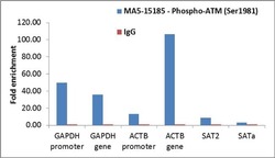
- Enrichment of endogenous Phospho-ATM (Ser1981) protein at specific gene loci using Anti-Phospho-ATM (Ser1981) Antibody: Chromatin Immunoprecipitation (ChIP) was performed using Anti-Phospho-ATM (Ser1981) Mouse Monoclonal Antibody (Product # MA5-15185, 6 µl) on sheared chromatin from 2 million HeLa cells treated with Etoposide (50 µM for 3 hours) using the MAGnify ChIP system kit (Product # 49-2024). Normal Rabbit IgG was used as a negative IP control. The purified DNA was analyzed by qPCR with PCR primer pairs over GAPDH and ACTB promoters and genes (active) and SAT2 satellite repeats, SAT alpha (inactive). Data is presented as fold enrichment of the antibody signal versus the negative control IgG using the comparative CT method.

- Submitted by
- Invitrogen Antibodies (provider)
- Main image

- Experimental details
- Enrichment of endogenous Phospho-ATM (Ser1981) protein at specific gene loci using Anti-Phospho-ATM (Ser1981) Antibody: Chromatin Immunoprecipitation (ChIP) was performed using Anti-Phospho-ATM (Ser1981) Mouse Monoclonal Antibody (Product # MA5-15185, 6 µl) on sheared chromatin from 2 million HeLa cells treated with Etoposide (50 µM for 3 hours) using the MAGnify ChIP system kit (Product # 49-2024). Normal Rabbit IgG was used as a negative IP control. The purified DNA was analyzed by qPCR with PCR primer pairs over GAPDH and ACTB promoters and genes (active) and SAT2 satellite repeats, SAT alpha (inactive). Data is presented as fold enrichment of the antibody signal versus the negative control IgG using the comparative CT method.
