 Explore
 Validate
 Learn
Learn Western blot
 Immunocytochemistry
Antibody data
- Antibody Data
- Antigen structure
- References [1]
- Comments [0]
- Validations
- Immunocytochemistry [2]
- Immunoprecipitation [1]
- Other assay [1]
Submit
Validation data
Reference
Comment
Report error
- Product number
- MA5-14366 - Provider product page

- Provider
- Invitrogen Antibodies
- Product name
- Cdk5 Monoclonal Antibody (DC34)
- Antibody type
- Monoclonal
- Antigen
- Recombinant full-length protein
- Description
- MA5-14366 targets Cdk5 in immunofluorescence, immunoprecipitation, and Western blot applications and shows reactivity with human, mouse, and rat samples. The MA5-14366 immunogen is recombinant cdk5 protein.
- Reactivity
- Human, Mouse, Rat
- Host
- Mouse
- Isotype
- IgG
- Antibody clone number
- DC34
- Vial size
- 500 μL
- Concentration
- 0.2 mg/mL
- Storage
- 4°C
Submitted references (+/-)-huprine Y, (-)-huperzine A and tacrine do not show neuroprotective properties in an apoptotic model of neuronal cytoskeletal alteration.
Jordá EG, Verdaguer E, Jiménez A, Canudas AM, Rimbau V, Camps P, Muñoz-Torrero D, Camins A, Pallàs M
Journal of Alzheimer's disease : JAD 2004 Dec;6(6):577-83; discussion 673-81
Journal of Alzheimer's disease : JAD 2004 Dec;6(6):577-83; discussion 673-81
No comments: Submit comment
Supportive validation
- Submitted by
- Invitrogen Antibodies (provider)
- Main image
- Experimental details
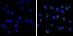
- Immunofluorescent analysis of Cdk5 (green) showing staining in the cytoplasm of Jurkat cells (right) compared to a negative control without primary antibody (left). Formalin-fixed cells were permeabilized with 0.1% Triton X-100 in TBS for 5-10 minutes and blocked with 3% BSA-PBS for 30 minutes at room temperature. Cells were probed with a Cdk5 monoclonal antibody (Product # MA5-14366) in 3% BSA-PBS at a dilution of 1:20 and incubated overnight at 4ºC in a humidified chamber. Cells were washed with PBST and incubated with a DyLight-conjugated secondary antibody in PBS at room temperature in the dark. F-actin (red) was stained with a fluorescent red phalloidin and nuclei (blue) were stained with Hoechst or DAPI. Images were taken at a magnification of 60x.

- Submitted by
- Invitrogen Antibodies (provider)
- Main image
- Experimental details
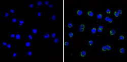
- Immunofluorescent analysis of Cdk5 (green) showing staining in the cytoplasm of Jurkat cells (right) compared to a negative control without primary antibody (left). Formalin-fixed cells were permeabilized with 0.1% Triton X-100 in TBS for 5-10 minutes and blocked with 3% BSA-PBS for 30 minutes at room temperature. Cells were probed with a Cdk5 monoclonal antibody (Product # MA5-14366) in 3% BSA-PBS at a dilution of 1:20 and incubated overnight at 4ºC in a humidified chamber. Cells were washed with PBST and incubated with a DyLight-conjugated secondary antibody in PBS at room temperature in the dark. F-actin (red) was stained with a fluorescent red phalloidin and nuclei (blue) were stained with Hoechst or DAPI. Images were taken at a magnification of 60x.

Supportive validation
- Submitted by
- Invitrogen Antibodies (provider)
- Main image
- Experimental details
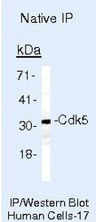
- Immunoprecipitation of Cdk5 using Cdk5 Monoclonal Antibody (MA5-14366) on Native Human LS174T Cells.

Supportive validation
- Submitted by
- Invitrogen Antibodies (provider)
- Main image
- Experimental details
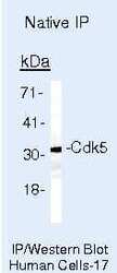
- Immunoprecipitation of Cdk5 using Cdk5 Monoclonal Antibody (MA5-14366) on Native Human LS174T Cells.
