 Explore
 Validate
 Learn
LearnPA5-29932
antibody from Invitrogen Antibodies
Targeting: ATP6AP2
APT6M8-9, ATP6IP2, ATP6M8-9, M8-9, PRR, RENR
 Western blot
Antibody data
- Antibody Data
- Antigen structure
- References [0]
- Comments [0]
- Validations
- Western blot [3]
- Immunocytochemistry [5]
Submit
Validation data
Reference
Comment
Report error
- Product number
- PA5-29932 - Provider product page

- Provider
- Invitrogen Antibodies
- Product name
- ATP6IP2 Polyclonal Antibody
- Antibody type
- Polyclonal
- Antigen
- Recombinant full-length protein
- Description
- Recommended positive controls: 293T, A431, H1299, HeLa, HepG2, Molt-4, Raji. Predicted reactivity: Mouse (92%), Rat (91%), Rhesus Monkey (98%), Bovine (87%). Store product as a concentrated solution. Centrifuge briefly prior to opening the vial.
- Reactivity
- Human, Mouse
- Host
- Rabbit
- Isotype
- IgG
- Vial size
- 100 μL
- Concentration
- 1.38 mg/mL
- Storage
- Store at 4°C short term. For long term storage, store at -20°C, avoiding freeze/thaw cycles.
No comments: Submit comment
Supportive validation
- Submitted by
- Invitrogen Antibodies (provider)
- Main image
- Experimental details
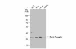
- Western Blot using ATP6IP2 Polyclonal Antibody (Product # PA5-29932). Various whole cell extracts (30 µg) were separated by 10% SDS-PAGE, and the membrane was blotted with ATP6IP2 Polyclonal Antibody (Product # PA5-29932) diluted at 1:1,000. The HRP-conjugated anti-rabbit IgG antibody was used to detect the primary antibody.

- Submitted by
- Invitrogen Antibodies (provider)
- Main image
- Experimental details
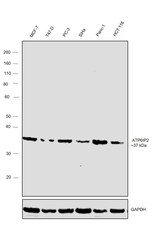
- Western blot was performed using Anti-ATP6IP2 Polyclonal Antibody (Product # PA5-29932) and a ~37 kD band was observed across the panel tested. Whole cell extracts (30 µg lysate) of MCF-7 (Lane 1), T-47D (Lane 2), PC-3 (Lane 3), SiHa (Lane 4), Panc-1 (Lane 5) and HCT 116 (Lane 6) were electrophoresed using NuPAGE™ 10% Bis-Tris Protein Gel (Product # NP0302BOX). Resolved proteins were then transferred onto a nitrocellulose membrane (Product # IB23001) by iBlot® 2 Dry Blotting System (Product # IB21001). The blot was probed with the primary antibody (1:1000 dilution) and detected by chemiluminescence with Goat anti-Rabbit IgG (Heavy Chain), Superclonal™ Recombinant Secondary Antibody, HRP (Product # A27036, 1:4000 dilution) using the iBright FL 1000 (Product # A32752). Chemiluminescent detection was performed using Novex® ECL Chemiluminescent Substrate Reagent Kit (Product # WP20005).

- Submitted by
- Invitrogen Antibodies (provider)
- Main image
- Experimental details
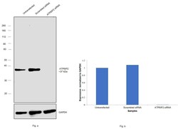
- Knockdown of ATP6IP2 was achieved by transfecting MCF-7 with ATP6IP2 specific siRNAs (Silencer® select Product # s19791, 19790). Western blot analysis (Fig. a) was performed using whole cell extracts from the ATP6IP2 knockdown cells (lane 3), non-specific scrambled siRNA transfected cells (lane 2) and untransfected cells (lane 1). The blot was probed with Arp3 Polyclonal Antibody (Product # PA5-29932, 1:1,000 dilution) and Goat anti-Rabbit IgG (Heavy Chain) Superclonal™ Recombinant Secondary Antibody, HRP (Product # A27036, 0.25 µg/mL, 1:4,000 dilution). Densitometric analysis of this western blot is shown in histogram (Fig. b). Decrease in signal upon siRNA mediated knock down confirms that antibody is specific to ATP6IP2.

Supportive validation
- Submitted by
- Invitrogen Antibodies (provider)
- Main image
- Experimental details
- Immunofluorescent analysis of Renin Receptor in methanol-fixed HepG2 cells using a Renin Receptor polyclonal antibody (Product # PA5-29932) at a 1:200 dilution.

- Submitted by
- Invitrogen Antibodies (provider)
- Main image
- Experimental details
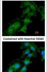
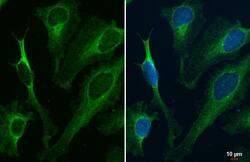
- Renin Receptor antibody detects Renin Receptor protein at endoplasmic reticulum by immunofluorescent analysis. Sample: HeLa cells were fixed in ice-cold MeOH for 5 min. Green: Renin Receptor stained by Renin Receptor antibody (Product # PA5-29932) diluted at 1:500.

- Submitted by
- Invitrogen Antibodies (provider)
- Main image

- Experimental details
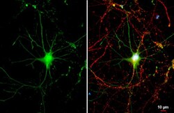
- ATP6IP2 Polyclonal Antibody detects Renin Receptor protein by immunofluorescent analysis. Sample: DIV9 rat hippocampal neuron cells were fixed in 4% paraformaldehyde at RT for 15 min. Green: Renin Receptor stained by ATP6IP2 Polyclonal Antibody (Product # PA5-29932) diluted at 1:500. Red: Tau, a Axon marker, stained by Tau antibody [GT287] diluted at 1:500. Blue: Fluoroshield with DAPI .

- Submitted by
- Invitrogen Antibodies (provider)
- Main image

- Experimental details
- Renin Receptor antibody detects Renin Receptor protein at endoplasmic reticulum by immunofluorescent analysis. Sample: HeLa cells were fixed in ice-cold MeOH for 5 min. Green: Renin Receptor stained by Renin Receptor antibody (Product # PA5-29932) diluted at 1:500.

- Submitted by
- Invitrogen Antibodies (provider)
- Main image
- Experimental details
- ATP6IP2 Polyclonal Antibody detects Renin Receptor protein by immunofluorescent analysis. Sample: DIV9 rat hippocampal neuron cells were fixed in 4% paraformaldehyde at RT for 15 min. Green: Renin Receptor stained by ATP6IP2 Polyclonal Antibody (Product # PA5-29932) diluted at 1:500. Red: Tau, a Axon marker, stained by Tau antibody [GT287] diluted at 1:500. Blue: Fluoroshield with DAPI .
