 Explore
 Validate
 Learn
LearnPA5-25788
antibody from Invitrogen Antibodies
Targeting: CUX1
CASP, CDP, CDP/Cut, CDP/Cux, CDP1, Clox, CUT, CUTL1, CUX, Cux/CDP, GOLIM6
 Western blot
Antibody data
- Antibody Data
- Antigen structure
- References [0]
- Comments [0]
- Validations
- Western blot [3]
- Immunocytochemistry [2]
- Immunohistochemistry [4]
Submit
Validation data
Reference
Comment
Report error
- Product number
- PA5-25788 - Provider product page

- Provider
- Invitrogen Antibodies
- Product name
- CUX1 Polyclonal Antibody
- Antibody type
- Polyclonal
- Antigen
- Synthetic peptide
- Reactivity
- Human
- Host
- Rabbit
- Isotype
- IgG
- Vial size
- 400 μL
- Concentration
- 1.8 mg/mL
- Storage
- Store at 4°C short term. For long term storage, store at -20°C, avoiding freeze/thaw cycles.
No comments: Submit comment
Supportive validation
- Submitted by
- Invitrogen Antibodies (provider)
- Main image
- Experimental details
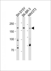
- Western blot analysis of CUX1 in various lysates. Samples were incubated with CUX1 polyclonal antibody (Product # PA5-25788) using a dilution of 1:1,000 followed by Goat Anti-Rabbit IgG, (H+L), Peroxidase conjugated at a dilution of 1:15,000. Lysates/proteins: 20 µg per lane. Lane 1: SH-SY5Y whole cell lysate; Lane 2: SK-BR-3 whole cell lysate; Lane 3: NIH/3T3 whole cell lysate. Observed band size: 200 kDa. Blocking/Dilution buffer: 5% NFDM/TBST.

- Submitted by
- Invitrogen Antibodies (provider)
- Main image
- Experimental details
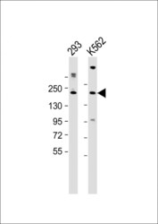
- Western blot analysis of CUX1 in various lysates. Samples were incubated with CUX1 polyclonal antibody (Product # PA5-25788) using a dilution of 1:2,000 followed by Goat Anti-Rabbit IgG, (H+L), Peroxidase conjugated at a dilution of 1:10,000. Lysates/proteins: 20 µg per lane. Lane 1: 293 whole cell lysate; Lane 2: K562 whole cell lysate. Predicted band size: 164 kDa. Blocking/Dilution buffer: 5% NFDM/TBST.

- Submitted by
- Invitrogen Antibodies (provider)
- Main image
- Experimental details
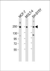
- Western blot analysis of CUX1 in various lysates. Samples were incubated with CUX1 polyclonal antibody (Product # PA5-25788) using a dilution of 1:2,000 followed by Goat Anti-Rabbit IgG, (H+L), Peroxidase conjugated at a dilution of 1:10,000. Lysates/proteins: 20 µg per lane. Lane 1: MCF-7 whole cell lysate; Lane 2: MOLT-4 whole cell lysate; Lane 3: SH-SY5Y whole cell lysate. Predicted band size: 164 kDa. Blocking/Dilution buffer: 5% NFDM/TBST.

Supportive validation
- Submitted by
- Invitrogen Antibodies (provider)
- Main image
- Experimental details
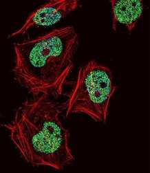
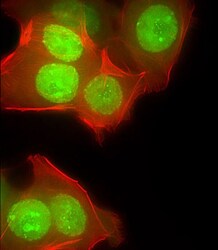
- Immunofluorescent analysis of HeLa cells using a CUX1 polyclonal antibody (Product # PA5-25788). HeLa cells were fixed with 4% PFA (20 min), permeabilized with Triton X-100 (0.1%, 10 min), then incubated with a CUX1 polyclonal antibody (Product # PA5-25788) (1:25, 1 hr at 37°C). Primary antibody was detected with fluor-conjugated donkey anti-rabbit secondary antibody (green) at 1:400 dilution for 50 min at 37°C). Actin filaments have been labeled with dye-conjugated phalloidin (red). Nuclei were counterstained with DAPI (blue) (10 µg/mL, 10 min).

- Submitted by
- Invitrogen Antibodies (provider)
- Main image
- Experimental details
- Immunocytochemistry analysis of CUX1 in MCF-7 cells. Samples were incubated with CUX1 polyclonal antibody (Product # PA5-25788) using a dilution of 1:25 followed by Dylight® 488-conjugated goat anti-rabbit IgG at a dilution of 1:200 (green). Cells were 4% paraformaldehyde-fixed and 0.1% Triton X-100 permeabilized. Immunofluorescence image showing nucleus and weak cytoplasm staining on MCF-7 cell line. Cytoplasmic actin is detected with Dylight® 554 Phalloidin at 1:100 dilution (red).

Supportive validation
- Submitted by
- Invitrogen Antibodies (provider)
- Main image

- Experimental details
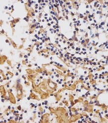
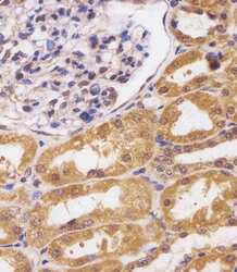
- Immunohistochemistry analysis of CUX1 in paraformaldehyde-fixed, paraffin-embedded human kidney tissue sections. Samples were incubated with CUX1 polyclonal antibody (Product # PA5-25788) using a dilution of 1:25 for 1 hours at 37°C followed by an undiluted biotinylated goat polyvalent antibody. Tissue was fixed with formaldehyde and blocked with 3% BSA for 0.5 hour at room temperature; antigen retrieval was by heat mediation with a citrate buffer (pH 6).

- Submitted by
- Invitrogen Antibodies (provider)
- Main image
- Experimental details
- Immunohistochemistry analysis of CUX1 in paraformaldehyde-fixed, paraffin-embedded human lymph node tissue sections. Samples were incubated with CUX1 polyclonal antibody (Product # PA5-25788) using a dilution of 1:25 for 1 hours at 37°C followed by an undiluted biotinylated goat polyvalent antibody. Tissue was fixed with formaldehyde and blocked with 3% BSA for 0.5 hour at room temperature; antigen retrieval was by heat mediation with a citrate buffer (pH 6).

- Submitted by
- Invitrogen Antibodies (provider)
- Main image
- Experimental details
- Immunohistochemistry analysis of CUX1 in paraformaldehyde-fixed, paraffin-embedded human kidney tissue sections. Samples were incubated with CUX1 polyclonal antibody (Product # PA5-25788) using a dilution of 1:25 for 1 hours at 37°C followed by an undiluted biotinylated goat polyvalent antibody. Tissue was fixed with formaldehyde and blocked with 3% BSA for 0.5 hour at room temperature; antigen retrieval was by heat mediation with a citrate buffer (pH 6).

- Submitted by
- Invitrogen Antibodies (provider)
- Main image

- Experimental details
- Immunohistochemistry analysis of CUX1 in paraformaldehyde-fixed, paraffin-embedded human lymph node tissue sections. Samples were incubated with CUX1 polyclonal antibody (Product # PA5-25788) using a dilution of 1:25 for 1 hours at 37°C followed by an undiluted biotinylated goat polyvalent antibody. Tissue was fixed with formaldehyde and blocked with 3% BSA for 0.5 hour at room temperature; antigen retrieval was by heat mediation with a citrate buffer (pH 6).
