 Explore
 Validate
 Learn
LearnPA5-30003
antibody from Invitrogen Antibodies
Targeting: CUX1
CASP, CDP, CDP/Cut, CDP/Cux, CDP1, Clox, CUT, CUTL1, CUX, Cux/CDP, GOLIM6
 Western blot
 Immunocytochemistry
Antibody data
- Antibody Data
- Antigen structure
- References [1]
- Comments [0]
- Validations
- Immunocytochemistry [4]
- Immunoprecipitation [1]
- Other assay [4]
Submit
Validation data
Reference
Comment
Report error
- Product number
- PA5-30003 - Provider product page

- Provider
- Invitrogen Antibodies
- Product name
- CUX1/Protein CASP Polyclonal Antibody
- Antibody type
- Polyclonal
- Antigen
- Recombinant full-length protein
- Description
- Recommended positive controls: Raji, K562. Predicted reactivity: Human (99%), Mouse (87%). Store product as a concentrated solution. Centrifuge briefly prior to opening the vial.
- Reactivity
- Human, Rat
- Host
- Rabbit
- Isotype
- IgG
- Vial size
- 100 μL
- Concentration
- 1 mg/mL
- Storage
- Store at 4°C short term. For long term storage, store at -20°C, avoiding freeze/thaw cycles.
Submitted references Overlapping Role of SCYL1 and SCYL3 in Maintaining Motor Neuron Viability.
Kuliyev E, Gingras S, Guy CS, Howell S, Vogel P, Pelletier S
The Journal of neuroscience : the official journal of the Society for Neuroscience 2018 Mar 7;38(10):2615-2630
The Journal of neuroscience : the official journal of the Society for Neuroscience 2018 Mar 7;38(10):2615-2630
No comments: Submit comment
Supportive validation
- Submitted by
- Invitrogen Antibodies (provider)
- Main image

- Experimental details
- CUX1/Protein CASP Polyclonal Antibody detects CUTL1 protein at Golgi apparatus by immunofluorescent analysis. Sample: SK-N-SH cells were fixed in 4% paraformaldehyde at RT for 15 min. Green: CUTL1 protein stained by CUX1/Protein CASP Polyclonal Antibody (Product # PA5-30003) diluted at 1:500. Blue: Hoechst 33342 staining. Scale bar = 10 µm.

- Submitted by
- Invitrogen Antibodies (provider)
- Main image

- Experimental details
- CUX1/Protein CASP Polyclonal Antibody detects CUTL1 protein by immunofluorescent analysis. Sample: DIV10 rat E18 primary cortical neuron cells were fixed in 4% paraformaldehyde at RT for 15 min. Green: CUTL1 stained by CUX1/Protein CASP Polyclonal Antibody (Product # PA5-30003) diluted at 1:500. Red: Tau, stained by Tau antibody [GT287] diluted at 1:500. Blue: Fluoroshield with DAPI .

- Submitted by
- Invitrogen Antibodies (provider)
- Main image

- Experimental details
- CUX1/Protein CASP Polyclonal Antibody detects CUTL1 protein at Golgi apparatus by immunofluorescent analysis. Sample: SK-N-SH cells were fixed in 4% paraformaldehyde at RT for 15 min. Green: CUTL1 protein stained by CUX1/Protein CASP Polyclonal Antibody (Product # PA5-30003) diluted at 1:500. Blue: Hoechst 33342 staining. Scale bar = 10 µm.

- Submitted by
- Invitrogen Antibodies (provider)
- Main image

- Experimental details
- CUX1/Protein CASP Polyclonal Antibody detects CUTL1 protein by immunofluorescent analysis. Sample: DIV10 rat E18 primary cortical neuron cells were fixed in 4% paraformaldehyde at RT for 15 min. Green: CUTL1 stained by CUX1/Protein CASP Polyclonal Antibody (Product # PA5-30003) diluted at 1:500. Red: Tau, stained by Tau antibody [GT287] diluted at 1:500. Blue: Fluoroshield with DAPI .

Supportive validation
- Submitted by
- Invitrogen Antibodies (provider)
- Main image

- Experimental details
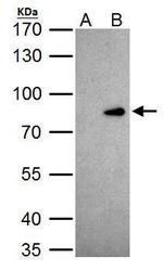
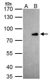
- CUX1/Protein CASP Polyclonal Antibody immunoprecipitates CUTL1 protein in IP experiments. IP Sample: 293T whole cell lysate/extract A. 40 µg 293T whole cell lysate/extract B. Control with 2 µg of preimmune rabbit IgG C. Immunoprecipitation of CUTL1 protein by 2 µg of CUX1/Protein CASP Polyclonal Antibody (Product # PA5-30003) 7.5% SDS-PAGE The immunoprecipitated CUTL1 protein was detected by CUX1/Protein CASP Polyclonal Antibody (Product # PA5-30003) diluted at 1:1,000.

Supportive validation
- Submitted by
- Invitrogen Antibodies (provider)
- Main image

- Experimental details
- NULL

- Submitted by
- Invitrogen Antibodies (provider)
- Main image

- Experimental details
- NULL

- Submitted by
- Invitrogen Antibodies (provider)
- Main image
- Experimental details
- CUX1/Protein CASP Polyclonal Antibody immunoprecipitates CUTL1 protein in IP experiments. IP Sample: 293T whole cell lysate/extract A. 40 µg 293T whole cell lysate/extract B. Control with 2 µg of preimmune rabbit IgG C. Immunoprecipitation of CUTL1 protein by 2 µg of CUX1/Protein CASP Polyclonal Antibody (Product # PA5-30003) 7.5% SDS-PAGE The immunoprecipitated CUTL1 protein was detected by CUX1/Protein CASP Polyclonal Antibody (Product # PA5-30003) diluted at 1:1,000.
- Submitted by
- Invitrogen Antibodies (provider)
- Main image

- Experimental details
- Figure 4. COPI and CASP interactions contribute to localization of SCYL3 to the Golgi apparatus. A , Generation of CASP-deficient MEFs using CRISPR-Cas9 technology. Structure of the Cux1 gene encoding CDP and CASP. Exons common to CDP and CASP are illustrated in black (only exons 12-14 illustrated). Exons encoding CDP are illustrated in green, and those encoding CASP are illustrated in blue. To generate CASP-deficient cells, 2 sgRNAs (black arrowheads) were designed to delete exons 15-17. Splicing of exon 14 into exon 18 causes a frame shift and premature stop codon. Red arrow, Cux1-F51 primer; green arrow, Cux-R32 primer. B , PCR genotyping of the WT and 2 independently derived CASP-deficient MEF lines (Cl.7 and Cl.8). Bands of 2496 and ~495 bp correspond to WT and CASP- alleles, respectively. A smaller band (*) from internal priming sites was also obtained by using these primers. NTC, No template control. Sanger sequencing confirmed proper recombination of the allele in CASP-deficient MEFs. C , Western blot analysis of CASP and beta-actin (as loading control) in WT and CASP-deficient MEFs. *Indicates nonspecific bands. D , Immunofluorescence staining of CASP and GM130 in WT and CASP-deficient MEFs. Exponentially growing WT and CASP-deficient MEFs were fixed and stained for CASP and GM130 and imaged by confocal microscopy. E , SCYL3 localization in WT and CASP-deficient MEFs. Exponentially growing WT and CASP-deficient MEFs were fixed and s
