Explore
 Validate
 Learn
Learn Western blot
Antibody data
- Antibody Data
- Antigen structure
- References [0]
- Comments [0]
- Validations
- Western blot [1]
- Immunohistochemistry [5]
- Flow cytometry [1]
Submit
Validation data
Reference
Comment
Report error
- Product number
- F52511 - Provider product page

- Provider
- NSJ Bioreagents
- Product name
- Histone H4 Antibody
- Antibody type
- Polyclonal
- Antigen
- This Histone H4 antibody was produced from a rabbit immunized with a KLH conjugated synthetic peptide between 71-103 amino acids from the C-terminal region of human HIST1H4A.
- Description
- Antigen affinity purified antibody
- Reactivity
- Human, Mouse, Rat
- Host
- Rabbit
- Conjugate
- Unconjugated
- Vial size
- 80 µl, 400 µl
- Storage
- Aliquot the Histone H4 antibody and store frozen at -20°C or colder. Avoid repeated freeze-thaw cycles.
No comments: Submit comment
Supportive validation
- Submitted by
- NSJ Bioreagents (provider)
- Main image
- Experimental details
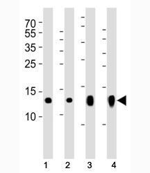
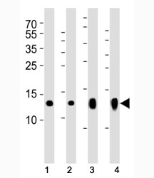
- Western blot analysis of lysate from (1) A431, (2) HeLa, (3) H-4-II-E and (4) L929 cell lines using Histone H4 antibody at 1:1000.
Supportive validation
- Submitted by
- NSJ Bioreagents (provider)
- Main image
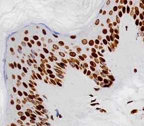
- Experimental details
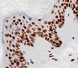
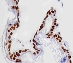
- Immunohistochemical analysis of paraffin-embedded human skin section using Histone H4 antibody diluted at 1:25 dilution.
- Submitted by
- NSJ Bioreagents (provider)
- Main image

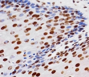
- Experimental details
- Immunohistochemical analysis of paraffin-embedded human esophagus section using Histone H4 antibody diluted at 1:25 dilution.
- Submitted by
- NSJ Bioreagents (provider)
- Main image
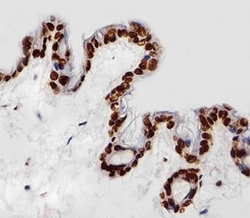
- Experimental details
- Immunohistochemical analysis of paraffin-embedded mouse skin section using Histone H4 antibody diluted at 1:25 dilution.

- Submitted by
- NSJ Bioreagents (provider)
- Main image

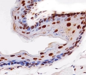
- Experimental details
- Immunohistochemical analysis of paraffin-embedded mouse esophagus section using Histone H4 antibody diluted at 1:25 dilution.
- Submitted by
- NSJ Bioreagents (provider)
- Main image
- Experimental details
- Immunohistochemical analysis of paraffin-embedded rat skin section using Histone H4 antibody diluted at 1:25 dilution.

Supportive validation
- Submitted by
- NSJ Bioreagents (provider)
- Main image
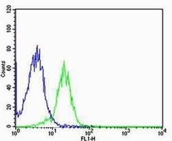
- Experimental details
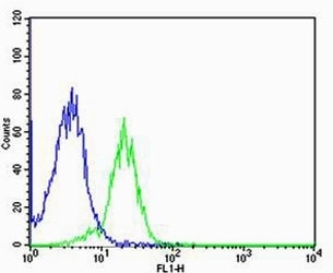
- Flow cytometric analysis of MCF-7 cells using Histone H4 antibody (green) compared to an isotype control of rabbit IgG (blue); Ab was diluted at 1:25 dilution. An Alexa Fluor 488 goat anti-rabbit lgG was used as the secondary Ab.