Explore
 Validate
 Learn
Learn Western blot
 Immunocytochemistry
 Immunoprecipitation
Antibody data
- Antibody Data
- Antigen structure
- References [2]
- Comments [0]
- Validations
- Immunocytochemistry [3]
- Other assay [3]
Submit
Validation data
Reference
Comment
Report error
- Product number
- PA5-17668 - Provider product page

- Provider
- Invitrogen Antibodies
- Product name
- Phospho-Glucocorticoid Receptor (Ser211) Polyclonal Antibody
- Antibody type
- Polyclonal
- Antigen
- Synthetic peptide
- Description
- It is not recommended to aliquot this antibody. This antibody is not cross-reactive with other unrelated phosphorylated proteins.
- Reactivity
- Human, Mouse
- Host
- Rabbit
- Isotype
- IgG
- Vial size
- 100 μL
- Concentration
- 74 μg/mL
- Storage
- -20°C
Submitted references Vitamin D interferes with glucocorticoid responsiveness in human peripheral blood mononuclear target cells.
BAY 11-7085 induces glucocorticoid receptor activation and autophagy that collaborate with apoptosis to induce human synovial fibroblast cell death.
Kassi E, Nasiri-Ansari N, Spilioti E, Kalotychou V, Apostolou PE, Moutsatsou P, Papavassiliou AG
Cellular and molecular life sciences : CMLS 2016 Nov;73(22):4341-4354
Cellular and molecular life sciences : CMLS 2016 Nov;73(22):4341-4354
BAY 11-7085 induces glucocorticoid receptor activation and autophagy that collaborate with apoptosis to induce human synovial fibroblast cell death.
Relic B, Charlier E, Deroyer C, Malaise O, Neuville S, Desoroux A, Gillet P, de Seny D, Malaise MG
Oncotarget 2016 Apr 26;7(17):23370-82
Oncotarget 2016 Apr 26;7(17):23370-82
No comments: Submit comment
Supportive validation
- Submitted by
- Invitrogen Antibodies (provider)
- Main image
- Experimental details
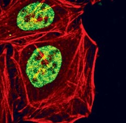
- Immunofluorescent analysis of Phospho-Glucocorticoid Receptor pSer211 in HeLa cells, dexamethasone-treated, using a Phospho-Glucocorticoid Receptor pSer211 polyclonal antibody (Product # PA5-17668) (green). Actin filaments are labeled with a fluorescent red phalloidin.
- Submitted by
- Invitrogen Antibodies (provider)
- Main image

- Experimental details
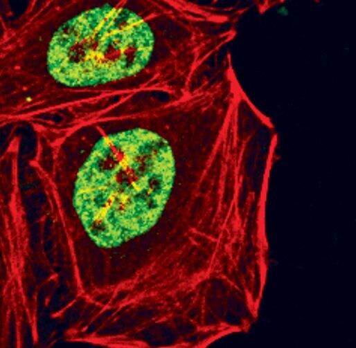
- Immunofluorescent analysis of Phospho-Glucocorticoid Receptor (Ser211) using a polyclonal antibody (Product # PA5-17668).

- Submitted by
- Invitrogen Antibodies (provider)
- Main image

- Experimental details
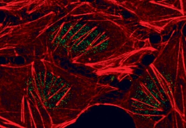
- Immunofluorescent analysis of Phospho-Glucocorticoid Receptor (Ser211) using a polyclonal antibody (Product # PA5-17668).
Supportive validation
- Submitted by
- Invitrogen Antibodies (provider)
- Main image
- Experimental details
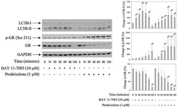
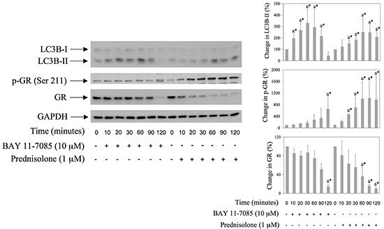
- Figure 1 BAY 11-7085 and prednisolone induce autophagy, GR phosphorylation and GR down regulation in human synovial fibroblasts Cells were cultured with BAY 11-7085 or prednisolone for the time indicated. Western blots show expression of p-GR (Serine 211), GR, LC3B and GAPDH in synovial fibroblast cell extracts. Graphs represent average of protein expression from three different experiments done with synovial fibroblasts from three different OA patients, expressed as a percentage of control. a* statistically different from control cells.
- Submitted by
- Invitrogen Antibodies (provider)
- Main image
- Experimental details
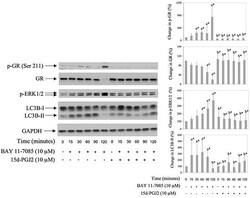
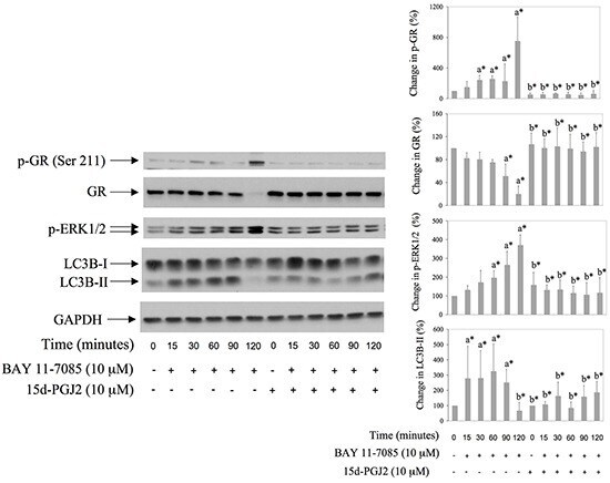
- Figure 3 BAY 11-7085 induced autophagy, GR phosphorylation on Serine 211 and GR degradation are inhibited with PPARgamma agonist 15d-PGJ2 Synovial fibroblasts were pretreated with 15d-PGJ2 for 24 hours and then BAY 11-7985 was added for the time indicated. Western blots show expression of p-GR (Ser 211), GR, p-ERK1/2, LC3B and GAPDH in synovial fibroblast cell extracts. Graphs represent average of protein expression from three different experiments done with synovial fibroblasts from three different OA patients, expressed as a percentage of control. a* statistically different from control cells; b* statistically different from a*.
- Submitted by
- Invitrogen Antibodies (provider)
- Main image

- Experimental details
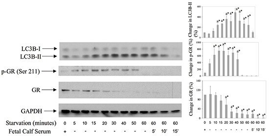
- Figure 6 GR is both phosphorylated on Serine 211 and down regulated during starvation-induced autophagy in synovial fibroblsts Cells were first starved by serum deprivation from the culture medium for 5-60 minutes, and then serum (10%) was replaced or not for additional 5-15 minutes. Western blots show expression of p-GR (Ser 211), GR, LC3B and GAPDH in synovial fibroblast cell extracts. Graphs represent average of protein expression from three different experiments done with synovial fibroblasts from three different OA patients, expressed as a percentage of control. a* statistically different from control cells.