Explore
 Validate
 Learn
Learn Western blot
Antibody data
- Antibody Data
- Antigen structure
- References [2]
- Comments [0]
- Validations
- Western blot [1]
- Immunohistochemistry [4]
- Flow cytometry [2]
- Other assay [1]
Submit
Validation data
Reference
Comment
Report error
- Product number
- PA5-14632 - Provider product page

- Provider
- Invitrogen Antibodies
- Product name
- ErbB2 (HER-2) Polyclonal Antibody
- Antibody type
- Polyclonal
- Antigen
- Synthetic peptide
- Description
- This antibody is predicted to react with mouse and rat based on sequence homology.
- Reactivity
- Human
- Host
- Rabbit
- Isotype
- IgG
- Vial size
- 400 μL
- Concentration
- 2 mg/mL
- Storage
- Store at 4°C short term. For long term storage, store at -20°C, avoiding freeze/thaw cycles.
Submitted references Th1 cytokines in conjunction with pharmacological Akt inhibition potentiate apoptosis of breast cancer cells in vitro and suppress tumor growth in vivo.
High-resolution clonal mapping of multi-organ metastasis in triple negative breast cancer.
Showalter L, Czerniecki BJ, Koski GK
Oncotarget 2020 Jul 28;11(30):2873-2888
Oncotarget 2020 Jul 28;11(30):2873-2888
High-resolution clonal mapping of multi-organ metastasis in triple negative breast cancer.
Echeverria GV, Powell E, Seth S, Ge Z, Carugo A, Bristow C, Peoples M, Robinson F, Qiu H, Shao J, Jeter-Jones SL, Zhang X, Ramamoorthy V, Cai S, Wu W, Draetta G, Moulder SL, Symmans WF, Chang JT, Heffernan TP, Piwnica-Worms H
Nature communications 2018 Nov 29;9(1):5079
Nature communications 2018 Nov 29;9(1):5079
No comments: Submit comment
Supportive validation
- Submitted by
- Invitrogen Antibodies (provider)
- Main image
- Experimental details
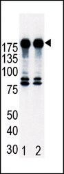
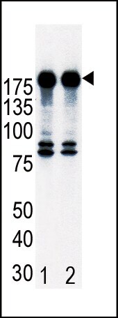
- Western blot analysis of ErbB2 (HER-2) in T47D cell lysates. Samples were incubated with ErbB2 (HER-2) polyclonal antibody (Product # PA5-14632). Lane 1: non-induced or Lane 2: induced with HRG.
Supportive validation
- Submitted by
- Invitrogen Antibodies (provider)
- Main image
- Experimental details
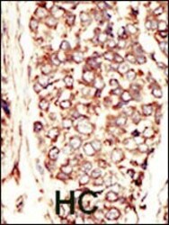
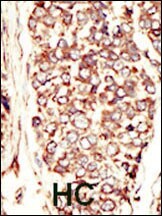
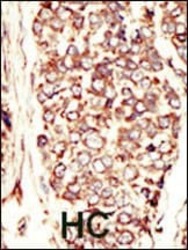
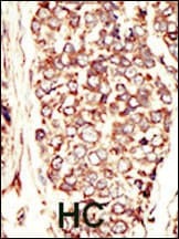
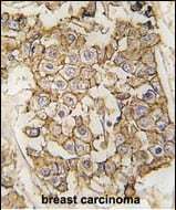
- Immunohistochemistry analysis of ErbB2 (HER-2) in formalin-fixed and paraffin-embedded human cancer tissue. Samples were incubated with ErbB2 (HER-2) polyclonal antibody (Product # PA5-14632) which was peroxidase-conjugated to the secondary antibody, followed by AEC staining. This data demonstrates the use of this antibody for immunohistochemistry; clinical relevance has not been evaluated. BC = breast carcinoma; HC = hepatocarcinoma.
- Submitted by
- Invitrogen Antibodies (provider)
- Main image
- Experimental details
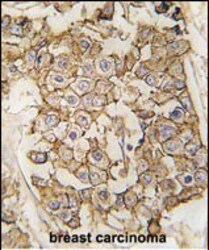
- Immunohistochemistry analysis of ErbB2 (HER-2) in formalin-fixed and paraffin-embedded human breast carcinoma tissue. Samples were incubated with ErbB2 (HER-2) polyclonal antibody (Product # PA5-14632) which was peroxidase-conjugated to the secondary antibody, followed by DAB staining. This data demonstrates the use of this antibody for immunohistochemistry; clinical relevance has not been evaluated.

- Submitted by
- Invitrogen Antibodies (provider)
- Main image
- Experimental details
- Immunohistochemistry analysis of ErbB2 (HER-2) in formalin-fixed and paraffin-embedded human cancer tissue. Samples were incubated with ErbB2 (HER-2) polyclonal antibody (Product # PA5-14632) which was peroxidase-conjugated to the secondary antibody, followed by AEC staining. This data demonstrates the use of this antibody for immunohistochemistry; clinical relevance has not been evaluated. BC = breast carcinoma; HC = hepatocarcinoma.
- Submitted by
- Invitrogen Antibodies (provider)
- Main image

- Experimental details
- Immunohistochemistry analysis of ErbB2 (HER-2) in formalin-fixed and paraffin-embedded human breast carcinoma tissue. Samples were incubated with ErbB2 (HER-2) polyclonal antibody (Product # PA5-14632) which was peroxidase-conjugated to the secondary antibody, followed by DAB staining. This data demonstrates the use of this antibody for immunohistochemistry; clinical relevance has not been evaluated.
Supportive validation
- Submitted by
- Invitrogen Antibodies (provider)
- Main image

- Experimental details
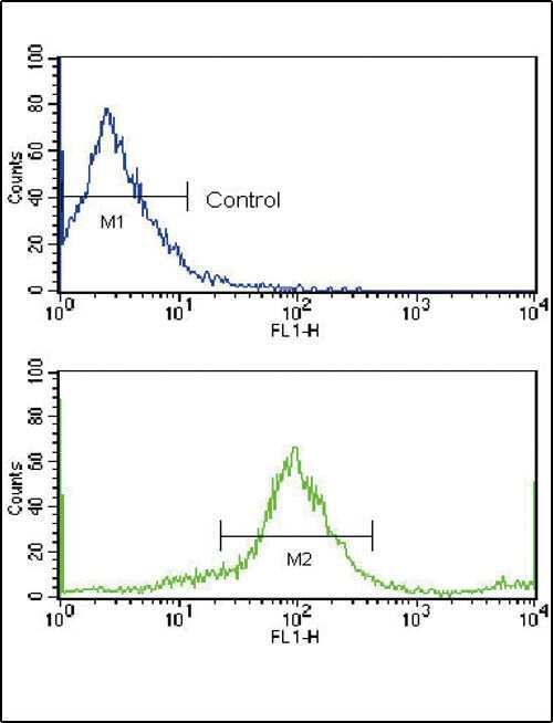
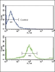
- Flow cytometry analysis of MCF-7 cells using a HER2/ErbB2 polyclonal antibody (Product # PA5-14632) (bottom), compared to a negative control cell (top) at a dilution of 1:10-50, followed by a FITC-conjugated goat anti-rabbit antibody
- Submitted by
- Invitrogen Antibodies (provider)
- Main image
- Experimental details
- Flow cytometry of ErbB2 (HER-2) in MCF-7 cells (bottom histogram). Samples were incubated with ErbB2 (HER-2) polyclonal antibody (Product # PA5-14632) followed by FITC-conjugated goat-anti-rabbit secondary antibody. Negative control (top histogram).

Supportive validation
- Submitted by
- Invitrogen Antibodies (provider)
- Main image

- Experimental details
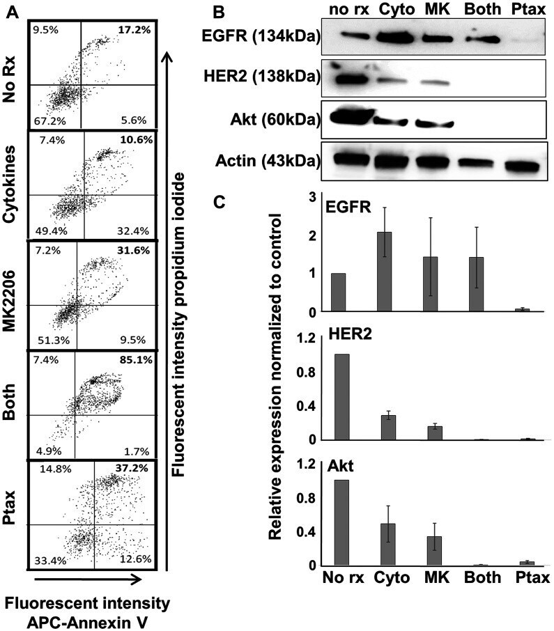
- Figure 5 Th1 cytokines, MK-2206, and paclitaxel induce apoptosis in human breast cancer cells but differentially affect expression of oncodrivers. SKBR3 cells were seeded at a density of 1 x 10 5 per well in 12-well cluster plates, and cultured overnight. The next day cells were exposed to Th1 cytokines (IFN-gamma and TNF-alpha at 10 ng/mL each), MK-2206 (10 muM), both treatments, paclitaxel (300 nM) or left untreated. After 72 h further incubation, cells were harvested and ( A ) stained with propidium iodide and APC-labeled Annexin V. Stained cells were subjected to flow cytometry and data evaluated by quadrant analysis, with double-staining cells (upper right quadrant) defined as apoptotic. ( B ) Western blot analysis of cell extracts for expression of EGFR, HER2, Akt kinase and actin (loading control). ( C ) Densitometry analysis of western blots (composite data from 3 separate experiments) normalized to actin expression. Error bars indicate SEM.