Explore
 Validate
 Learn
Learn Western blot
 Immunocytochemistry
Antibody data
- Antibody Data
- Antigen structure
- References [3]
- Comments [0]
- Validations
- Immunocytochemistry [2]
- Immunohistochemistry [1]
- Flow cytometry [2]
- Other assay [2]
Submit
Validation data
Reference
Comment
Report error
- Product number
- PA5-14635 - Provider product page

- Provider
- Invitrogen Antibodies
- Product name
- ErbB2 (HER-2) Polyclonal Antibody
- Antibody type
- Polyclonal
- Antigen
- Recombinant full-length protein
- Reactivity
- Human
- Host
- Rabbit
- Isotype
- IgG
- Vial size
- 400 μL
- Concentration
- 0.40 mg/mL
- Storage
- Store at 4°C short term. For long term storage, store at -20°C, avoiding freeze/thaw cycles.
Submitted references Downregulation of miR-375 contributes to ERBB2-mediated VEGFA overexpression in esophageal cancer.
Dasatinib inhibition of cSRC prevents the migration and metastasis of canine mammary cancer cells with enhanced Wnt and HER signalling.
High basal Wnt signaling is further induced by PI3K/mTor inhibition but sensitive to cSRC inhibition in mammary carcinoma cell lines with HER2/3 overexpression.
Ren S, Tan X, Fu MZ, Ren S, Wu X, Chen T, Latham PS, Lin P, Man YG, Fu SW
Journal of Cancer 2021;12(23):7138-7146
Journal of Cancer 2021;12(23):7138-7146
Dasatinib inhibition of cSRC prevents the migration and metastasis of canine mammary cancer cells with enhanced Wnt and HER signalling.
Timmermans-Sprang EPM, Mestemaker HM, Steenlage RR, Mol JA
Veterinary and comparative oncology 2019 Sep;17(3):413-426
Veterinary and comparative oncology 2019 Sep;17(3):413-426
High basal Wnt signaling is further induced by PI3K/mTor inhibition but sensitive to cSRC inhibition in mammary carcinoma cell lines with HER2/3 overexpression.
Timmermans-Sprang EP, Gracanin A, Mol JA
BMC cancer 2015 Jul 25;15:545
BMC cancer 2015 Jul 25;15:545
No comments: Submit comment
Supportive validation
- Submitted by
- Invitrogen Antibodies (provider)
- Main image
- Experimental details
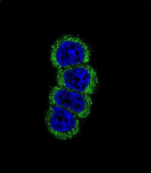
- Immunofluorescent analysis of MCF-7 cells using a HER2 polyclonal antibody (Product # PA5-14635) at a dilution of 1:10-50, followed by a fluor-conjugated goat anti-rabbit secondary antibody (green). Nuclei were stained with DAPI (blue).

- Submitted by
- Invitrogen Antibodies (provider)
- Main image

- Experimental details
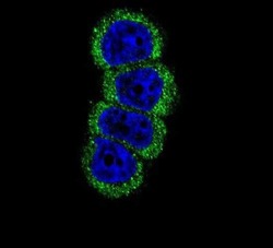
- Immunocytochemistry analysis of ErbB2 (HER-2) in MCF-7 cells. Samples were incubated in ErbB2 (HER-2) polyclonal antibody (Product # PA5-14635) followed by Alexa Fluor 488-conjugated goat anti-rabbit lgG (green). DAPI was used to stain the cell nuclear (blue).
Supportive validation
- Submitted by
- Invitrogen Antibodies (provider)
- Main image

- Experimental details
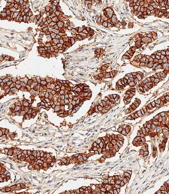
- Immunohistochemistry analysis of ErbB2 (HER-2) in paraffin-embedded Human breast carcinoma tissue. Samples were incubated with ErbB2 (HER-2) polyclonal antibody (Product # PA5-14635) using a dilution of 1:500 for 1 hour at room temperature followed by an undiluted biotinylated CRF Anti-Polyvalent HRP Polymer antibody. Tissue was fixed with formaldehyde at room temperature, antigen retrieval was by heat mediation with a EDTA buffer (pH 9.0).
Supportive validation
- Submitted by
- Invitrogen Antibodies (provider)
- Main image

- Experimental details
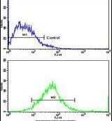
- Flow cytometry analysis of MCF-7 cells using a HER2 polyclonal antibody (Product # PA5-14635) (bottom), compared to a negative control cell (top) at a dilution of 1:10-50, followed by a FITC-conjugated goat anti-rabbit antibody
- Submitted by
- Invitrogen Antibodies (provider)
- Main image
- Experimental details
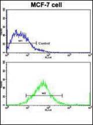
- Flow cytometry of ErbB2 (HER-2) in MCF-7 cells (bottom histogram). Samples were incubated with ErbB2 (HER-2) polyclonal antibody (Product # PA5-14635) followed by FITC-conjugated goat-anti-rabbit secondary antibody. Negative control cell (top histogram).

Supportive validation
- Submitted by
- Invitrogen Antibodies (provider)
- Main image
- Experimental details
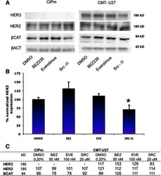
- Fig. 6 Protein levels in CIPm and CMT-U27 inhibited with BEZ235, Everolimus and Src-I1. Cells were cultured for 40 h with the inhibitors, 50nM BEZ235, 100nM Everolimus and 20 muM Src-l1. Total protein was isolated with RIPA buffer and 20 mug protein was used for Western Blot analyses. Blots were probed with total antibodies for beta-Catenin, HER2, HER3 and beta-Actin ( a ). The HER3 blot was done in triplicate and normalized against beta-Actin. Results, expressed as % of control are the mean (+-STDEV) * P < 0,05 versus DMSO control ( b ). Densities were measured, corrected for the background and related to beta-Actin expression as loading control, expressed in % ( c )

- Submitted by
- Invitrogen Antibodies (provider)
- Main image

- Experimental details
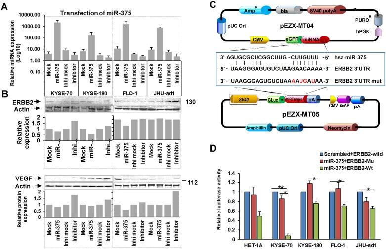
- Figure 4 miR-375 regulates ERBB2 and VEGFA in EC . A. Expression of miR-375 after transfection. B. Forced expression of miR-375 in EC cell lines resulted in decreased ERBB2 and VEGFA expression. C. Map of the plasmids pEZX-MT04 containing miR-375, and pEZX-MT05 containing 3'-UTR of ERBB2 to illustrate the binding site of miR-375 at the 3'-UTR of ERBB2, and its mutant control sequence. D. Dual luciferase reporter assay. Co-transfection with pEZX-miR-375 and pEZX-ERBB2 3' UTR wild type significantly decreased the luciferase activities compared to that with pEZX-miR-375 and ERBB2 3' UTR mutant/miR-375 scrambled control and ERBB2 3' UTR wild type sequence in ESCC cell lines. The data were reported as mean +- S.D. from three independent experiments (** p < 0.01, *p