 Explore
 Validate
 Learn
LearnPA3-16862
antibody from Invitrogen Antibodies
Targeting: CALR
cC1qR, CRT, FLJ26680, RO, SSA
 Western blot
 Immunocytochemistry Immunohistochemistry
 Blocking/Neutralizing Immunoelectron microscopy Other assay
Antibody data
- Antibody Data
- Antigen structure
- References [0]
- Comments [0]
- Validations
- Immunocytochemistry [3]
- Immunohistochemistry [2]
Submit
Validation data
Reference
Comment
Report error
- Product number
- PA3-16862 - Provider product page

- Provider
- Invitrogen Antibodies
- Product name
- Calreticulin Polyclonal Antibody
- Antibody type
- Polyclonal
- Antigen
- Other
- Description
- Suggested positive control: antigen standard for CALR (transient overexpression lysate).
- Reactivity
- Human, Mouse, Rat, Bovine, Hamster
- Host
- Rabbit
- Isotype
- IgG
- Vial size
- 100 μL
- Concentration
- 1 mg/mL
- Storage
- -20°C or -80°C if preferred
No comments: Submit comment
Supportive validation
- Submitted by
- Invitrogen Antibodies (provider)
- Main image

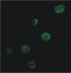
- Experimental details
- Immunofluorescence staining of Calreticulin in HCT15 colon cancer cells using Product # PA3-16862. Secondary antibody was an Alexa Fluor 488.
- Submitted by
- Invitrogen Antibodies (provider)
- Main image
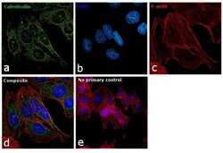
- Experimental details
- Immunofluorescence analysis of Calreticulin was performed using 70% confluent log phase U-2 OS cells. The cells were fixed with 4% paraformaldehyde for 10 minutes, permeabilized with 0.1% Triton™ X-100 for 15 minutes, and blocked with 1% BSA for 1 hour at room temperature. The cells were labeled with Calreticulin Polyclonal Antibody (Product # PA3-16862) at 1:200 dilution in 0.1% BSA, incubated at 4 degree Celsius overnight and then labeled with Goat anti-Rabbit IgG (H+L) Superclonal™ Secondary Antibody, Alexa Fluor® 488 conjugate (Product # A27034) at a dilution of 1:2000 for 45 minutes at room temperature (Panel a: green). Nuclei (Panel b: blue) were stained with SlowFade® Gold Antifade Mountant with DAPI (Product # S36938). F-actin (Panel c: red) was stained with Rhodamine Phalloidin (Product # R415, 1:300). Panel d represents the merged image showing endoplasmic reticulum and mitochondrial localization. Panel e represents control cells with no primary antibody to assess background. The images were captured at 60X magnification.
- Submitted by
- Invitrogen Antibodies (provider)
- Main image

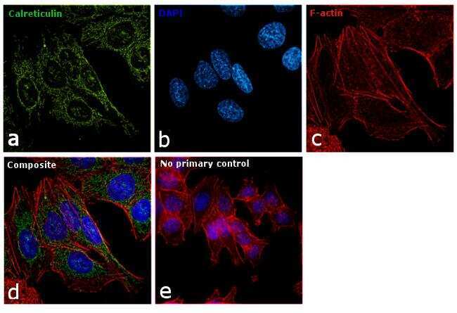
- Experimental details
- Immunofluorescence analysis of Calreticulin was performed using 70% confluent log phase U-2 OS cells. The cells were fixed with 4% paraformaldehyde for 10 minutes, permeabilized with 0.1% Triton™ X-100 for 15 minutes, and blocked with 1% BSA for 1 hour at room temperature. The cells were labeled with Calreticulin Polyclonal Antibody (Product # PA3-16862) at 1:200 dilution in 0.1% BSA, incubated at 4 degree Celsius overnight and then labeled with Goat anti-Rabbit IgG (Heavy Chain) Superclonal™ Secondary Antibody, Alexa Fluor® 488 conjugate (Product # A27034) at a dilution of 1:2000 for 45 minutes at room temperature (Panel a: green). Nuclei (Panel b: blue) were stained with SlowFade® Gold Antifade Mountant with DAPI (Product # S36938). F-actin (Panel c: red) was stained with Rhodamine Phalloidin (Product # R415, 1:300). Panel d represents the merged image showing endoplasmic reticulum and mitochondrial localization. Panel e represents control cells with no primary antibody to assess background. The images were captured at 60X magnification.

Supportive validation
- Submitted by
- Invitrogen Antibodies (provider)
- Main image

- Experimental details
- Immunohistochemical analysis of Calreticulin in formalin-fixed paraffin-embedded tissue section of human thyroid gland. Samples were incubated in Calreticulin polyclonal antibody (Product # PA3-16862) using a dilution of 1:50. The signal was developed using HRP-DAB based detection method which followed counterstaining of the nuclei with hematoxylin. This representative section shows a strong positivity of Calreticulin in the follicular epithelial cells, wherein the signal was found to be very intense in the perinuclear region of the cells which correlates well with Endoplasmic reticulum localization of this protein. The para-follicular cells, endothelial cells of blood vessels (not the RBCs though) and the loose connective tissue in the section showed a weak cytoplasmic staining. Some staining was observed in the follicles/colloids also which is potentially the secreted form of Calreticulin.

- Submitted by
- Invitrogen Antibodies (provider)
- Main image

- Experimental details
- Immunohistochemical analysis of Calreticulin in formalin-fixed paraffin-embedded tissue section of human thyroid gland. Samples were incubated in Calreticulin polyclonal antibody (Product # PA3-16862) using a dilution of 1:50. The signal was developed using HRP-DAB based detection method which followed counterstaining of the nuclei with hematoxylin. This representative section shows a strong positivity of Calreticulin in the follicular epithelial cells, wherein the signal was found to be very intense in the perinuclear region of the cells which correlates well with Endoplasmic reticulum localization of this protein. The para-follicular cells, endothelial cells and the loose connective tissue in the section showed a weak cytoplasmic staining. Some staining was observed in the follicles/colloids also which is potentially the secreted form of Calreticulin.
