 Explore
 Validate
 Learn
Learn Western blot
Antibody data
- Antibody Data
- Antigen structure
- References [1]
- Comments [0]
- Validations
- Western blot [3]
- Immunocytochemistry [4]
- Immunohistochemistry [1]
- Other assay [1]
Submit
Validation data
Reference
Comment
Report error
- Product number
- MA5-15768 - Provider product page

- Provider
- Invitrogen Antibodies
- Product name
- XBP1 Monoclonal Antibody (9B7E5)
- Antibody type
- Monoclonal
- Antigen
- Recombinant full-length protein
- Description
- A suggested positive control is XBP-1 recombinant protein.
- Reactivity
- Human, Mouse, Rat
- Host
- Mouse
- Isotype
- IgG
- Antibody clone number
- 9B7E5
- Vial size
- 100 µg
- Concentration
- 1 mg/mL
- Storage
- -20°C
Submitted references Nuclear factor-kappa B-dependent X-box binding protein 1 signalling promotes the proliferation of nucleus pulposus cells under tumour necrosis factor alpha stimulation.
Chen L, Xie ZY, Liu L, Zhu L, Wang F, Fan P, Sinkemani A, Zhang C, Hong X, Wu XT
Cell proliferation 2019 Mar;52(2):e12542
Cell proliferation 2019 Mar;52(2):e12542
No comments: Submit comment
Supportive validation
- Submitted by
- Invitrogen Antibodies (provider)
- Main image
- Experimental details
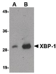
- Western blot analysis of 100 ng of XBP-1 recombinant protein using a XBP-1 monoclonal antibody (Product # MA5-15768) at 1 µg/mL.

- Submitted by
- Invitrogen Antibodies (provider)
- Main image
- Experimental details
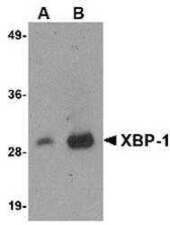
- Western Blot analysis of 100 ng of XBP-1 recombinant protein with XBP1 Monoclonal Antibody (9B7E5) (Product # MA5-15768) at 1 µg/mL.

- Submitted by
- Invitrogen Antibodies (provider)
- Main image

- Experimental details
- Western Blot analysis of 100 ng of XBP-1 recombinant protein with XBP1 Monoclonal Antibody (9B7E5) (Product # MA5-15768) at 1 µg/mL.

Supportive validation
- Submitted by
- Invitrogen Antibodies (provider)
- Main image

- Experimental details
- Immunofluorescent analysis of HepG2 cells using a XBP-1 monoclonal antibody (Product # MA5-15768) at a 2 µg/mL dilution.

- Submitted by
- Invitrogen Antibodies (provider)
- Main image

- Experimental details
- Immunocytochemistry of XBP-1 in HepG2 cells with XBP1 Monoclonal Antibody (9B7E5) (Product # MA5-15768) at 2 µg/mL.

- Submitted by
- Invitrogen Antibodies (provider)
- Main image

- Experimental details
- Immunofluorescence of XBP1 in HepG2 cells with XBP1 Monoclonal Antibody (9B7E5) (Product # MA5-15768) at 2 µg/mL.

- Submitted by
- Invitrogen Antibodies (provider)
- Main image

- Experimental details
- Immunofluorescence analysis of XBP1 was performed using 70% confluent log phase MCF7 thapsigargin treated cells (2uM for 6hrs). The cells were fixed with 4% paraformaldehyde for 10 minutes, permeabilized with 0.1% Triton™ X-100 for 15 minutes, and blocked with 2% BSA for 45 minutes at room temperature. The cells were labeled with XBP1 Monoclonal Antibody (9B7E5)(Product # MA5-15768) at 5µg/mL in 0.1% BSA, incubated at 4 degree celsius overnight and then labeled with Donkey anti-Mouse IgG (H+L) Highly Cross-Adsorbed Secondary Antibody, Alexa Fluor Plus 488 (Product # MA5-15768), (1:2000 dilution), for 45 minutes at room temperature (Panel a: Green). Nuclei (Panel b: Blue) were stained with ProLong™ Diamond Antifade Mountant with DAPI (Product # P36962) . F-actin (Panel c: Red) was stained with Rhodamine Phalloidin (Product # R415, 1:300 dilution). Panel d represents the merged image showing cytoplasmic localization. Panel e represents untreated cells with reduced signal. Panel f represents control cells with no primary antibody to assess background. The images were captured at 60X magnification.

Supportive validation
- Submitted by
- Invitrogen Antibodies (provider)
- Main image

- Experimental details
- Immunocytochemistry staining of HepG2 cells using a XBP-1 monoclonal antibody (Product # MA5-15768) at a 2 µg/mL dilution.

Supportive validation
- Submitted by
- Invitrogen Antibodies (provider)
- Main image

- Experimental details
- Figure 5 X-box binding protein 1 (XBP1) regulates nuclear factor-kappa B signalling by regulating p65 transcription and p -p65 expression. The cells were exposed to 10 ng/mL tumour necrosis factor alpha with or without pretreatment with siCtrl and siXBP1 for 24 h. A, In immunofluorescence double staining, the cells were stained initially with XBP1 antibody, Alexa Fluor 488 (green), p -p65 antibody and Alexa Fluor 647 (red), and the nucleus was counterstained with DAPI (blue) and examined through fluorescence microscopy (original magnification, x400). B, XBP1- and p -p65-positive cells were quantified. C, Total RNA was extracted, and the mRNA expression of p65 was measured through qRT-PCR. D,E, Proteins were extracted to detect the expression of p -p65 and p65 through Western blot analysis, and the protein expression levels of p -p65 were normalized to that of p65. The results were presented as mean +- SD ( n = 3). * P < 0.05; ** P < 0.01
