 Explore
 Validate
 Learn
Learn Western blot
Antibody data
- Antibody Data
- Antigen structure
- References [5]
- Comments [0]
- Validations
- Western blot [1]
- Flow cytometry [1]
- Chromatin Immunoprecipitation [1]
- Other assay [1]
Submit
Validation data
Reference
Comment
Report error
- Product number
- MA5-11319 - Provider product page

- Provider
- Invitrogen Antibodies
- Product name
- E2F3 Monoclonal Antibody (3E2F04 (PG37))
- Antibody type
- Monoclonal
- Antigen
- Recombinant full-length protein
- Description
- MA5-11319 targets E2F-3 Transcription Factor in GS and IF applications and shows reactivity with Human samples.
- Antibody clone number
- 3E2F04 (PG37)
- Concentration
- 0.2 mg/mL
Submitted references Chromatin remodeling protein HELLS is upregulated by inactivation of the RB-E2F pathway and is nonessential for osteosarcoma tumorigenesis.
Integrated genomic and gene expression profiling identifies two major genomic circuits in urothelial carcinoma.
Anti-apoptotic function of the E2F transcription factor 4 (E2F4)/p130, a member of retinoblastoma gene family in cardiac myocytes.
Inactivation of the Rb pathway and overexpression of both isoforms of E2F3 are obligate events in bladder tumours with 6p22 amplification.
E2F3 amplification and overexpression is associated with invasive tumor growth and rapid tumor cell proliferation in urinary bladder cancer.
Wu SC, Benavente CA
Oncotarget 2018 Aug 24;9(66):32580-32592
Oncotarget 2018 Aug 24;9(66):32580-32592
Integrated genomic and gene expression profiling identifies two major genomic circuits in urothelial carcinoma.
Lindgren D, Sjödahl G, Lauss M, Staaf J, Chebil G, Lövgren K, Gudjonsson S, Liedberg F, Patschan O, Månsson W, Fernö M, Höglund M
PloS one 2012;7(6):e38863
PloS one 2012;7(6):e38863
Anti-apoptotic function of the E2F transcription factor 4 (E2F4)/p130, a member of retinoblastoma gene family in cardiac myocytes.
Dingar D, Konecny F, Zou J, Sun X, von Harsdorf R
Journal of molecular and cellular cardiology 2012 Dec;53(6):820-8
Journal of molecular and cellular cardiology 2012 Dec;53(6):820-8
Inactivation of the Rb pathway and overexpression of both isoforms of E2F3 are obligate events in bladder tumours with 6p22 amplification.
Hurst CD, Tomlinson DC, Williams SV, Platt FM, Knowles MA
Oncogene 2008 Apr 24;27(19):2716-27
Oncogene 2008 Apr 24;27(19):2716-27
E2F3 amplification and overexpression is associated with invasive tumor growth and rapid tumor cell proliferation in urinary bladder cancer.
Oeggerli M, Tomovska S, Schraml P, Calvano-Forte D, Schafroth S, Simon R, Gasser T, Mihatsch MJ, Sauter G
Oncogene 2004 Jul 22;23(33):5616-23
Oncogene 2004 Jul 22;23(33):5616-23
No comments: Submit comment
Supportive validation
- Submitted by
- Invitrogen Antibodies (provider)
- Main image

- Experimental details
- Western blot analysis was performed on whole cell extracts (30 µg lysate) of HeLa (Lane 1), SH-SY5Y (lane 2), U-87MG (lane 3), MDA-MB-231 (Lane 4), PC-3 (lane 5). The blots were probed with Anti-E2F3 Transcription Factor Mouse Monoclonal Antibody (Product # MA5-11319, 2 µg/mL) and detected by chemiluminescence Goat anti-Mouse IgG (H+L) Secondary Antibody, HRP conjugate (Product # 62-6520, 1:4000 dilution). Two bands 37 and 49 kDa band corresponding to E2F3 Transcription Factor was observed across cell lines tested. Known quantity of protein samples were electrophoresed using Novex® NuPAGE® 10 % Bis-Tris gel (Product # NP0301BOX), XCell SureLock™ Electrophoresis System (Product # EI0002) and Novex® Sharp Pre-Stained Protein Standard (Product # LC5800). Resolved proteins were then transferred onto a nitrocellulose membrane with iBlot® 2 Dry Blotting System (Product # IB21001). The membrane was probed with the relevant primary and secondary Antibody following blocking with 5 % skimmed milk. Chemiluminescent detection was performed using Pierce™ ECL Western Blotting Substrate (Product # 32106).

Supportive validation
- Submitted by
- Invitrogen Antibodies (provider)
- Main image
- Experimental details
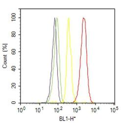
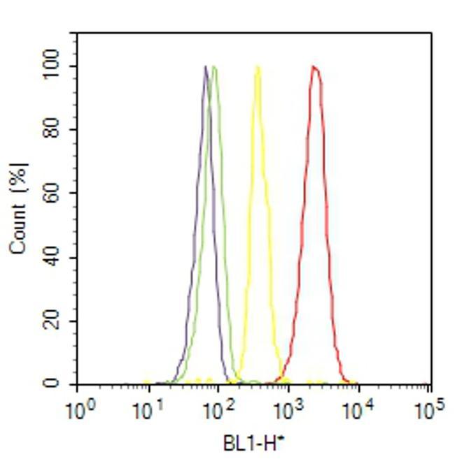
- Flow cytometry analysis of E2F-3 Transcription Factor was done on K562 cells. Cells were fixed with 70% ethanol for 10 minutes, permeabilized with 0.25% Triton™ X-100 for 20 minutes, and blocked with 5% BSA for 30 minutes at room temperature. Cells were labeled with E2F-3 Transcription Factor Mouse Monoclonal Antibody (MA511319, red histogram) or with mouse isotype control (yellow histogram) at 3-5 ug/million cells in 2.5% BSA. After incubation at room temperature for 2 hours, the cells were labeled with Alexa Fluor® 488 Rabbit Anti-Mouse Secondary Antibody (A11059) at a dilution of 1:400 for 30 minutes at room temperature. The representative 10,000 cells were acquired and analyzed for each sample using an Attune® Acoustic Focusing Cytometer. The purple histogram represents unstained control cells and the green histogram represents no-primary-antibody control..
Supportive validation
- Submitted by
- Invitrogen Antibodies (provider)
- Main image

- Experimental details
- Enrichment of endogenous E2F3 Protein at specific gene loci using Anti-E2F3 Transcription Factor Mouse Monoclonal Antibody: Chromatin Immunoprecipitation (ChIP) was performed using Anti-E2F3 Transcription Factor Mouse Monoclonal Antibody (Product # MA5-11319, 5 µg) on sheared chromatin from 2 million K562 cells using the "MAGnify ChIP system" kit (Product # 49-2024). Normal Mouse IgG was used as a negative IP control. The purified DNA was analyzed by 7500 Fast qPCR system (Product # 4351106) with optimized PCR primer pairs for the promoter of active E2F3, ATATD2, RAD51 gene, used as positive control target, and the SAT2, used as negative control target. Data is presented as fold enrichment of the antibody signal versus the negative control IgG using the comparative CT method.

Supportive validation
- Submitted by
- Invitrogen Antibodies (provider)
- Main image

- Experimental details
- Figure 3 HELLS is a direct transcriptional target of the RB-E2F pathway ( A ) Chromatin immunoprecipitation (ChIP) assay reveals enrichment of E2F1 within HELLS promoter. Primers flanking the myoglobin promoter were used as negative control. Each point is mean +- s.d. of triplicate samples. ( B - C ) Western blot detection of HELLS protein level in osteosarcoma cells transduced with lentivirus containing shRNA against (B) E2F1 (shE2F1) show no significant change in HELLS expression, but ( C ) combination of E2F1 (shE2F1) and E2F3 (shE2F3) knockdown decreased HELLS protein levels. ( D - E ) RT-qPCR analysis of basal expression of (D) CDK4 and (E) CDK6 mRNA in different osteosarcoma cell lines normalized to mesenchymal stem cells (MSC). Each point is mean +- s.d. of triplicate samples. ( F ) RT-qPCR analysis of HELLS mRNA expression in osteosarcoma cell lines after Palbociclib-treated for 24 h, normalized to DMSO control cells. Each point is mean +- s.d. of triplicate samples. * p < 0.0332, ** p < 0.0021, *** p < 0.0002, **** p < 0.0001 by two-tailed t test.
