 Explore
 Validate
 Learn
Learn Western blot
 ELISA
ELISAAntibody data
- Antibody Data
- Antigen structure
- References [8]
- Comments [0]
- Validations
- Western blot [2]
- Immunocytochemistry [1]
- Other assay [6]
Submit
Validation data
Reference
Comment
Report error
- Product number
- MA1-83428 - Provider product page

- Provider
- Invitrogen Antibodies
- Product name
- PGP9.5 Monoclonal Antibody (31A3)
- Antibody type
- Monoclonal
- Antigen
- Other
- Description
- This antibody does not react with guinea pig. This antibody is suitable for use on paraffin-embedded tissue sections, with a recommend fixation in 95% ethanol/5% acetic acid for 2-3 hours prior to paraffin-embedding. Specimens which have not been fixed in acetic acid/alcohol will likely require pretreatment using the microwave-citrate buffer method. Mouse anti Human Protein Gene Product 9.5 antibody, clone 31A3 recognizes protein gene product 9.5 (PGP9.5), a ubiquitin hydrolase which is widely expressed in neuronal tissues and represents 1-2% of total soluble brain proteins.
- Reactivity
- Human, Mouse, Rat, Rabbit
- Host
- Mouse
- Isotype
- IgG
- Antibody clone number
- 31A3
- Vial size
- 200 µg
- Concentration
- 1 mg/mL
- Storage
- Store at 4°C short term. For long term storage, store at -20°C, avoiding freeze/thaw cycles.
Submitted references The ephrin receptor EphB2 regulates the connectivity and activity of enteric neurons.
Repurposing mechanistic insight of PDE-5 inhibitor in cancer chemoprevention through mitochondrial-oxidative stress intervention and blockade of DuCLOX signalling.
Activation of prolyl hydroxylase-2 for stabilization of mitochondrial stress along with simultaneous downregulation of HIF-1α/FASN in ER + breast cancer subtype.
Chemical activation of prolyl hydroxylase-2 by BBAP-1 down regulates hypoxia inducible factor-1α and fatty acid synthase for mammary gland chemoprevention.
Alpha-linolenic acid stabilizes HIF-1 α and downregulates FASN to promote mitochondrial apoptosis for mammary gland chemoprevention.
Comparative efficacy of alpha-linolenic acid and gamma-linolenic acid to attenuate valproic acid-induced autism-like features.
α-Chymotrypsin regulates free fatty acids and UCHL-1 to ameliorate N-methyl nitrosourea induced mammary gland carcinoma in albino wistar rats.
Effect of β-sitosterol against methyl nitrosourea-induced mammary gland carcinoma in albino rats.
Bodin R, Paillé V, Oullier T, Durand T, Aubert P, Le Berre-Scoul C, Hulin P, Neunlist M, Cissé M
The Journal of biological chemistry 2021 Nov;297(5):101300
The Journal of biological chemistry 2021 Nov;297(5):101300
Repurposing mechanistic insight of PDE-5 inhibitor in cancer chemoprevention through mitochondrial-oxidative stress intervention and blockade of DuCLOX signalling.
Singh M, Kasna S, Roy S, Aldosary S, Saeedan AS, Ansari MN, Kaithwas G
BMC cancer 2019 Oct 24;19(1):996
BMC cancer 2019 Oct 24;19(1):996
Activation of prolyl hydroxylase-2 for stabilization of mitochondrial stress along with simultaneous downregulation of HIF-1α/FASN in ER + breast cancer subtype.
Devi U, Singh M, Roy S, Gupta PS, Ansari MN, Saeedan AS, Kaithwas G
Cell biochemistry and function 2019 Jun;37(4):216-227
Cell biochemistry and function 2019 Jun;37(4):216-227
Chemical activation of prolyl hydroxylase-2 by BBAP-1 down regulates hypoxia inducible factor-1α and fatty acid synthase for mammary gland chemoprevention.
Singh M, Devi U, Roy S, Gupta PS, Kaithwas G
RSC advances 2018 Apr 3;8(23):12848-12860
RSC advances 2018 Apr 3;8(23):12848-12860
Alpha-linolenic acid stabilizes HIF-1 α and downregulates FASN to promote mitochondrial apoptosis for mammary gland chemoprevention.
Roy S, Rawat AK, Sammi SR, Devi U, Singh M, Gautam S, Yadav RK, Rawat JK, Singh L, Ansari MN, Saeedan AS, Pandey R, Kumar D, Kaithwas G
Oncotarget 2017 Sep 19;8(41):70049-70071
Oncotarget 2017 Sep 19;8(41):70049-70071
Comparative efficacy of alpha-linolenic acid and gamma-linolenic acid to attenuate valproic acid-induced autism-like features.
Yadav S, Tiwari V, Singh M, Yadav RK, Roy S, Devi U, Gautam S, Rawat JK, Ansari MN, Saeedan AS, Prakash A, Saraf SA, Kaithwas G
Journal of physiology and biochemistry 2017 May;73(2):187-198
Journal of physiology and biochemistry 2017 May;73(2):187-198
α-Chymotrypsin regulates free fatty acids and UCHL-1 to ameliorate N-methyl nitrosourea induced mammary gland carcinoma in albino wistar rats.
Rani A, Roy S, Singh M, Devi U, Yadav RK, Gautam S, Rawat JK, Ansari MN, Saeedan AS, Prakash A, Kaithwas G
Inflammopharmacology 2016 Oct;24(5):277-286
Inflammopharmacology 2016 Oct;24(5):277-286
Effect of β-sitosterol against methyl nitrosourea-induced mammary gland carcinoma in albino rats.
Manral C, Roy S, Singh M, Gautam S, Yadav RK, Rawat JK, Devi U, Ansari MN, Saeedan AS, Kaithwas G
BMC complementary and alternative medicine 2016 Jul 29;16:260
BMC complementary and alternative medicine 2016 Jul 29;16:260
No comments: Submit comment
Supportive validation
- Submitted by
- Invitrogen Antibodies (provider)
- Main image
- Experimental details
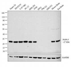
- Western blot analysis was performed on whole cell extracts (30 µg lysate) of Neuro-2a (Lane 1), SH-SY5Y (Lane 2), U-87 MG (Lane 3), PC-12 (Lane 4), HEK-293 (Lane 5), A549 (Lane 6), HeLa (Lane 7), Hep G2 (Lane 8), NIH/3T3 (Lane 9), tissue extracts of Mouse Brain (Lane 10) and Rat Brain (Lane 11). The blot was probed with Anti-PGP9.5 Monoclonal Antibody (Product # MA1-83428, 1µg/ml) and detected by chemiluminescence using Goat anti-Mouse IgG (H+L) Superclonal™ Secondary Antibody, HRP conjugate (Product # A28177, 0.25 µg/ml, 1:4000 dilution). A 27 kDa band corresponding to PGP9.5 was detected across the cell lines and tissues tested except for Hela, HepG2 and NIH/3T3 which is reported to be negative for PGP9.5 expression.

- Submitted by
- Invitrogen Antibodies (provider)
- Main image

- Experimental details
- Western blot analysis of PGP9.5/UCHL1 was performed by loading 20 µg of SH-SY5Y wild type (Lane 1), SH-SY5Y Cas9 control (Lane 2), SH-SY5Y PGP9.5/UCHL1 knockout (Lane 3) whole cell extracts. The blot was probed with Anti-PGP9.5/ UCHL1 Monoclonal Antibody (Product # MA1-83428) (1 µg/mL) and Goat anti-Mouse IgG (H+L), Superclonal™ Recombinant Secondary Antibody, HRP (Product # A28177) (1:4000 dilution). Loss of signal upon CRISPR mediated knockout (KO) confirms that antibody is specific to PGP9.5/UCHL1.

Supportive validation
- Submitted by
- Invitrogen Antibodies (provider)
- Main image

- Experimental details
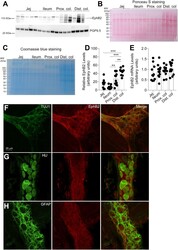
- Immunofluorescence analysis of PGP9.5 was performed using 70% confluent log phase SH-SY5Y cells. The cells were fixed with 4% paraformaldehyde for 10 minutes, permeabilized with 0.1% Triton™ X-100 for 10 minutes, and blocked with 1% BSA for 1 hour at room temperature. The cells were labeled with PGP9.5 Monoclonal Antibody (31A3) (Product # MA1-83428) at 5 µg/mL in 0.1% BSA and incubated overnight at 4 degree and then labeled with Goat anti-Mouse IgG (H+L) Superclonal™ Secondary Antibody, Alexa Fluor® 488 conjugate (Product # A28175) at a dilution of 1:2000 for 45 minutes at room temperature (Panel a: green). Nuclei (Panel b: blue) were stained with SlowFade® Gold Antifade Mountant with DAPI (Product # S36938). F-actin (Panel c: red) was stained with Rhodamine Phalloidin (Product # R415, 1:300). Panel d represents the merged image showing cytoplasmic localization. Panel e represents control cells with no primary antibody to assess background. The images were captured at 60X magnification.

Supportive validation
- Submitted by
- Invitrogen Antibodies (provider)
- Main image

- Experimental details
- NULL

- Submitted by
- Invitrogen Antibodies (provider)
- Main image

- Experimental details
- Fig. 4 Western Blot analysis: Group I: Control; Group II: MNU; Group III: beta-sitosterol10 mg/kg; Group IV: beta-sitosterol 20 mg/kg. The western blot analysis containing whole tissue lysate from rat mammary gland was probed for P-gp 9.5 antibody; its immunoreactivity was evident as a band of molecular weight of 27 kDa. In case of control the expression is low in comparison with the toxicant. The low and high dose of beta-sitosterol shows a similar pattern of elevated expression in a dose dependent manner. Results of the beta-actin analysis are shown as an internal control

- Submitted by
- Invitrogen Antibodies (provider)
- Main image
- Experimental details
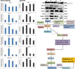
- 5 Downregulation of hypoxic pathway in mammary gland cancer. Immunoblotting of respective individual group (group 1--control [0.9% normal saline, p.o. ]; group 2--toxic control [MNU, 8 mg/kg, i.v. ]; group 3--BBAP-2 + MNU [68.52 mug/kg, s.c. + 8 mg/kg MNU, i.v. ]; and group 4--BBAP-2 + MNU [137.04 mug/kg, s.c. + 8 mg/kg MNU, i.v.]) was performed for estimation of HIF-1alpha, PHD2, and FASN as well as PHD2 in tumour growth. Perspective correlations of NFkappaBp65, UCHL-1, and SREBP-1c with the above-mentioned protein was well defined through the experimental pathway. Excised mammary gland tissue sample lyzed in trizol for RNA extraction and analysed for the mRNA expression of HIF-1alpha, PHD2, FASN, NFkappaBp65, UCHL-1, and SREBP-1c by real-time quantitative reverse transcription polymerase chain reaction (qRT-PCR): fold induction is relative to tissue under hypoxic conditions after normalization to the beta-actin expression. Columns: mean of three experimental determination; bars, SD. Comparisons are made on the basis of the one-way ANOVA followed by Bonferroni multiple test. All groups are compared with the toxic control group (* p< 0.05, ** p< 0.01, *** p< 0.001)

- Submitted by
- Invitrogen Antibodies (provider)
- Main image
- Experimental details
- Figure 1 EphB2 expression in different segments of the rat GI tract. A - C , EphB2 expression was examined by western blot analysis of full-thickness tissue sections from indicated regions of the rat GI tract. Pgp9.5 was used as a loading control ( A ). Note that pgp9.5 is expressed at higher levels in the distal colon. Furthermore, we observed the apparition of putative EphB2-derived bands in the colon that might be degradation products. Ponceau S ( B ) and Coomassie ( C ) stainings are provided as addition to the traditional loading control. Although equal amount of proteins was loaded for analysis, those stainings show the variability in protein migration profil and relative protein abundance between intestinal segments. Each data point represents the relative adjusted volume of protein band intensity measured by using a rectangle tool of constant volume on each lane of the WB membrane. Data are represented as relative values of protein density in arbitrary units. D , densitometric quantitation of western blot signals revealed that EphB2 is present in all segments of the gut (n = 12 sections per segment from 12 rats; jej., jejunum; prox. colon, proximal colon; dist. colon, distal colon; n = 12 sections per segment from 12 animals, F (3, 44) = 18.91, p = 0.00000005, ANOVA; Tukey's post-hoc test; ** p < 0.001, *** p < 0.0001). E , quantitative RT-PCR analysis of EphB2 mRNA levels in full-thickness tissue sections in the GI tract of adult rats. Note that there is no strict pr

- Submitted by
- Invitrogen Antibodies (provider)
- Main image

- Experimental details
- Effect of BBAP-1 treatment on hypoxic pathway in mammary gland cancer: protein was extracted from individual groups [1: control; 2: toxic control; 3: BBAP-1 + MNU (61.55 mug kg -1 s.c. + 8 mg kg -1 i.v. ); 4: BBAP-1 + MNU (123.11 mug kg -1 s.c. + 8 mg kg -1 i.v.)] and subjected to immunoblotting of PHD-2, HIF-1alpha, FASN and other successive markers (NFkappaBp65, UCHL-1 and SREBP-1c). The up-regulated expression of PHD-2 and down-regulated HIF-1alpha expression demonstrate the activation of PHD-2 by BBAP-1 treatment. Subsequent reduced expression of FASN validates the triggering of PHD-2 by BBAP-1. beta-actin was used as a loading control. Experimental values represent data derived from three individual experiments and are presented as mean +- SD. Comparisons are made on the basis of one-way ANOVA followed by Bonferroni multiple test. All groups are compared to the toxic control group (* p < 0.05, ** p < 0.01, *** p < 0.001).

- Submitted by
- Invitrogen Antibodies (provider)
- Main image

- Experimental details
- Fig. 8 Tadalafil mediated activation of mitochondrial mediated apoptosis pathway. Protein extracted from individual groups [Control (Normal saline, 3 ml/kg, p.o.); Toxic control (MNU 47 mg/kg, i.v.); MNU + Tadalafil (47 mg/kg i.v. + 2 mg/kg p.o.) and MNU + Tadalafil (47 mg/kg i.v. + 4 mg/kg p.o.)] were subjected to immunoblotting of proapoptotic (BAD) and anti-apoptotic (Bcl-xl) marker along with COX, NFkappaBp65 and UCHL-1. beta-actin was used as internal loading control. Each experiment was performed in triplicate. The data was represented as mean +- SD. The groups were significantly different by one-way ANOVA followed by Bonferroni multiple tests. All groups were compared to the toxic control group (* p < 0.05, ** p < 0.01, *** p < 0.001)
