 Explore
 Validate
 Learn
Learn Western blot
Antibody data
- Antibody Data
- Antigen structure
- References [1]
- Comments [0]
- Validations
- Western blot [3]
- Immunocytochemistry [3]
- Immunohistochemistry [3]
Submit
Validation data
Reference
Comment
Report error
- Product number
- PA1-20158 - Provider product page

- Provider
- Invitrogen Antibodies
- Product name
- PGP9.5 Polyclonal Antibody
- Antibody type
- Polyclonal
- Antigen
- Synthetic peptide
- Description
- Recommended positive controls: Human brain lysate. Store product as a concentrated solution. Centrifuge briefly prior to opening the vial.
- Reactivity
- Human, Mouse, Rat, Porcine
- Host
- Rabbit
- Isotype
- IgG
- Vial size
- 50 µL
- Concentration
- Conc. Not Determined
- Storage
- Store at 4°C short term. For long term storage, store at -20°C, avoiding freeze/thaw cycles.
Submitted references Local insulin and the rapid regrowth of diabetic epidermal axons.
Guo G, Kan M, Martinez JA, Zochodne DW
Neurobiology of disease 2011 Aug;43(2):414-21
Neurobiology of disease 2011 Aug;43(2):414-21
No comments: Submit comment
Supportive validation
- Submitted by
- Invitrogen Antibodies (provider)
- Main image
- Experimental details
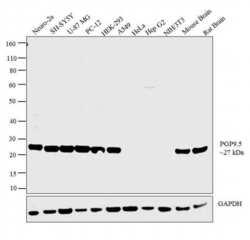
- Western blot analysis was performed on whole cell extracts (30 µg lysate) of Neuro-2a (Lane 1), SH-SY5Y (Lane 2), U-87 MG (Lane 3), PC-12 (Lane 4), HEK-293 (Lane 5), A549 (Lane 6), HeLa (Lane 7), Hep G2 (Lane 8), NIH/3T3 (Lane 9), tissue extracts of Mouse Brain (Lane 10) and Rat Brain (Lane 11). The blot was probed with Anti-PGP9.5 Polyclonal Antibody (Product # PA1-20158, 1:1000 dilution) and detected by chemiluminescence using Goat anti-Rabbit IgG (H+L) Superclonal™ Secondary Antibody, HRP conjugate (Product # A27036, 0.25 µg/ml, 1:4000 dilution). A 27 kDa band corresponding to PGP9.5 was detected across the cell lines and tissues tested except for Hela, HepG2 and NIH/3T3 which is reported to be negative for PGP9.5 expression.

- Submitted by
- Invitrogen Antibodies (provider)
- Main image
- Experimental details
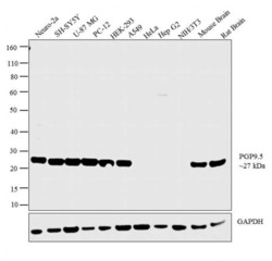
- Western blot analysis was performed on whole cell extracts (30 µg lysate) of Neuro-2a (Lane 1), SH-SY5Y (Lane 2), U-87 MG (Lane 3), PC-12 (Lane 4), HEK-293 (Lane 5), A549 (Lane 6), HeLa (Lane 7), Hep G2 (Lane 8), NIH/3T3 (Lane 9), tissue extracts of Mouse Brain (Lane 10) and Rat Brain (Lane 11). The blot was probed with Anti-PGP9.5 Polyclonal Antibody (Product # PA1-20158, 1:1000 dilution) and detected by chemiluminescence using Goat anti-Rabbit IgG (H+L) Superclonal™ Secondary Antibody, HRP conjugate (Product # A27036, 0.25 µg/ml, 1:4000 dilution). A 27 kDa band corresponding to PGP9.5 was detected across the cell lines and tissues tested except for Hela, HepG2 and NIH/3T3 which is reported to be negative for PGP9.5 expression.

- Submitted by
- Invitrogen Antibodies (provider)
- Main image
- Experimental details
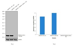
- Western blot analysis of PGP9.5/UCHL1 was performed by loading 20 µg of SHSY5Y wild type (Lane 1), SHSY5Y Cas9 control (Lane 2), SHSY5Y PGP9.5/ UCHL1 knockout (Lane 3) whole cell extracts. The blot was probed with Anti-PGP9.5 Polyclonal Antibody (Product # PA1-20158) (1:5000 dilution) and Goat anti-Mouse IgG (H+L), Superclonal™ Recombinant Secondary Antibody, HRP (Product # A28177) (1:4000 dilution). Loss of signal upon CRISPR mediated knockout (KO) confirms that antibody is specific to PGP9.5/ UCHL1.

Supportive validation
- Submitted by
- Invitrogen Antibodies (provider)
- Main image

- Experimental details
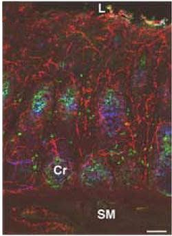
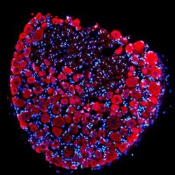
- Immunofluorescent analysis of PGP9.5 in porcine distal colon using a PGP9.5 polyclonal antibody (Product # PA1-20158) (red). Abbreviations: Cr, colonic crypt; L, intestinal lumen; SM, submucosa. Scale bars: 100µm.
- Submitted by
- Invitrogen Antibodies (provider)
- Main image

- Experimental details
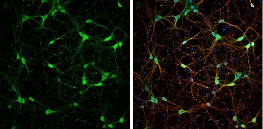
- Immunocytochemistry-Immunofluorescence analysis of PGP9.5 was performed in DIV9 rat E18 primary cortical neurons fixed in 4% paraformaldehyde at RT for 15 min. Green: PGP9.5 Polyclonal Antibody (Product # PA1-20158) diluted at 1:500. Red: beta Tubulin 3/ Tuj1, stained by beta Tubulin 3/ Tuj1 antibody. Blue: Fluoroshield with DAPI.
- Submitted by
- Invitrogen Antibodies (provider)
- Main image

- Experimental details
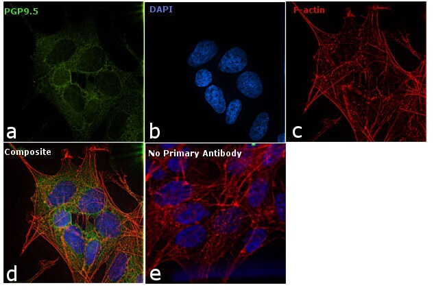
- Immunofluorescence analysis of PGP9.5 was performed using 70% confluent log phase SH-SY5Y cells. The cells were fixed with 4% paraformaldehyde for 10 minutes, permeabilized with 0.1% Triton™ X-100 for 10 minutes, and blocked with 1% BSA for 1 hour at room temperature. The cells were labeled with PGP9.5 Rabbit Polyclonal Antibody (Product # PA1-20158) at 1:250 dilution in 0.1% BSA and incubated overnight at 4 degree and then labeled with Goat anti-Mouse IgG (H+L) Superclonal™ Secondary Antibody, Alexa Fluor® 488 conjugate (Product # A27034) at a dilution of 1:2000 for 45 minutes at room temperature (Panel a: green). Nuclei (Panel b: blue) were stained with SlowFade® Gold Antifade Mountant with DAPI (Product # S36938). F-actin (Panel c: red) was stained with Rhodamine Phalloidin (Product # R415, 1:300). Panel d represents the merged image showing and cytoplasmic localization. Panel e represents control cells with no primary antibody to assess background. The images were captured at 60X magnification.
Supportive validation
- Submitted by
- Invitrogen Antibodies (provider)
- Main image
- Experimental details
- Immunohistochemistry analysis of rat DRG tissue using PGP9.5 Polyclonal Antibody (Product # PA1-20158). Dilution: 1:500.

- Submitted by
- Invitrogen Antibodies (provider)
- Main image
- Experimental details
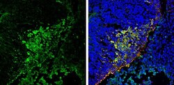
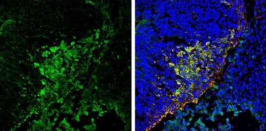
- Immunohistochemistry (Frozen) analysis of PGP9.5 was performed in frozen sectioned E13.5 Rat brain tissue using PGP9.5 Polyclonal Antibody (Product # PA1-20158) at a dilution of 1:250 (Green). Red: beta Tubulin 3/ TUJ1, a mature neuron marker, stained by beta Tubulin 3/ TUJ1 antibody diluted at 1:500. Blue: Fluoroshield with DAPI.
- Submitted by
- Invitrogen Antibodies (provider)
- Main image
- Experimental details
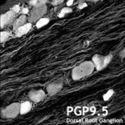
- Immunohistochemistry (Frozen) analysis of rat dorsal root ganglion using (Product # PA1-20158) PGP9.5 Polyclonal Antibody.
