Explore
 Validate
 Learn
Learn Western blot
 Immunocytochemistry
Antibody data
- Antibody Data
- Antigen structure
- References [3]
- Comments [0]
- Validations
- Western blot [9]
- Immunocytochemistry [3]
- Immunohistochemistry [5]
Submit
Validation data
Reference
Comment
Report error
- Product number
- GTX109637 - Provider product page

- Provider
- GeneTex
- Proper citation
- GeneTex Cat#GTX109637, RRID:AB_1952497
- Product name
- PGP9.5 antibody
- Antibody type
- Polyclonal
- Reactivity
- Human, Mouse, Rat
- Host
- Rabbit
Submitted references High-dose intravenous immunoglobulins reduce nerve macrophage infiltration and the severity of bortezomib-induced peripheral neurotoxicity in rats.
Na+ /K+ -ATPase coupled to endothelin receptor type B stimulates peripheral nerve regeneration via lactate signalling.
Hypo-osmotic shock-induced subclinical inflammation of skin in a rat model of disrupted skin barrier function.
Meregalli C, Marjanovic I, Scali C, Monza L, Spinoni N, Galliani C, Brivio R, Chiorazzi A, Ballarini E, Rodriguez-Menendez V, Carozzi VA, Alberti P, Fumagalli G, Pozzi E, Canta A, Quartu M, Briani C, Oggioni N, Marmiroli P, Cavaletti G
Journal of neuroinflammation 2018 Aug 21;15(1):232
Journal of neuroinflammation 2018 Aug 21;15(1):232
Na+ /K+ -ATPase coupled to endothelin receptor type B stimulates peripheral nerve regeneration via lactate signalling.
Tu NH, Katano T, Matsumura S, Funatsu N, Pham VM, Fujisawa JI, Ito S
The European journal of neuroscience 2017 Sep;46(5):2096-2107
The European journal of neuroscience 2017 Sep;46(5):2096-2107
Hypo-osmotic shock-induced subclinical inflammation of skin in a rat model of disrupted skin barrier function.
Kishi C, Minematsu T, Huang L, Mugita Y, Kitamura A, Nakagami G, Yamane T, Yoshida M, Noguchi H, Funakubo M, Mori T, Sanada H
Biological research for nursing 2015 Mar;17(2):135-41
Biological research for nursing 2015 Mar;17(2):135-41
No comments: Submit comment
Enhanced validation
Supportive validation
- Submitted by
- GeneTex (provider)
- Enhanced method
- Genetic validation
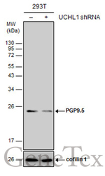
- Main image
- Experimental details
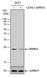
- Non-transfected (¡V) and transfected (+) 293T whole cell extracts (30 ?g) were separated by 12% SDS-PAGE, and the membrane was blotted with PGP9.5 antibody (GTX109637) diluted at 1:2000. The HRP-conjugated anti-rabbit IgG antibody (GTX213110-01) was used to detect the primary antibody.

Supportive validation
- Submitted by
- GeneTex (provider)
- Main image

- Experimental details
- Sample (30 ug of whole cell lysate) A: A549 B: H1299 12% SDS PAGE GTX109637 diluted at 1:5000
- Validation comment
- WB
- Submitted by
- GeneTex (provider)
- Main image
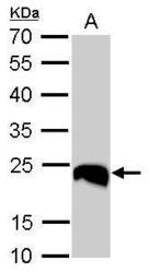
- Experimental details
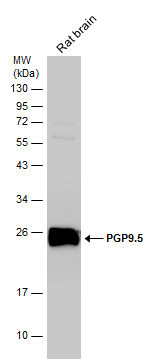
- PGP9.5 antibody detects UCHL1 protein by western blot analysis.A. 50 ?g mouse brain lysate/extract12% SDS-PAGEPGP9.5 antibody (GTX109637) dilution: 1:5000 The HRP-conjugated anti-rabbit IgG antibody (GTX213110-01) was used to detect the primary antibody.

- Submitted by
- GeneTex (provider)
- Main image
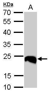
- Experimental details
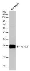
- PGP9.5 antibody detects UCHL1 protein by Western blot analysis.A. 50 µg rat brain lysate/extract12 % SDS-PAGEPGP9.5 antibody (GTX109637) dilution: 1:10000
- Validation comment
- WB
- Submitted by
- GeneTex (provider)
- Main image

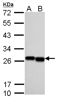
- Experimental details
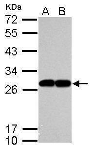
- PGP9.5 antibody detects PGP9.5 protein by western blot analysis.A. 30 ?g A549 whole cell lysate/extractB. 30 ?g H1299 whole cell lysate/extract12% SDS-PAGEPGP9.5 antibody (GTX109637) dilution: 1:10000The HRP-conjugated anti-rabbit IgG antibody (GTX213110-01) was used to detect the primary antibody.
- Submitted by
- GeneTex (provider)
- Main image
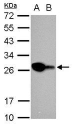
- Experimental details
- PGP9.5 antibody detects PGP9.5 protein by western blot analysis.A. 30 ?g Neuro2A whole cell lysate/extractB. 30 ?g GL261 whole cell lysate/extract12% SDS-PAGEPGP9.5 antibody (GTX109637) dilution: 1:10000The HRP-conjugated anti-rabbit IgG antibody (GTX213110-01) was used to detect the primary antibody.

- Submitted by
- GeneTex (provider)
- Main image
- Experimental details
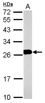
- Rat tissue extract (50 ?g) was separated by 12% SDS-PAGE, and the membrane was blotted with PGP9.5 antibody (GTX109637) diluted at 1:10000. The HRP-conjugated anti-rabbit IgG antibody (GTX213110-01) was used to detect the primary antibody.
- Submitted by
- GeneTex (provider)
- Main image
- Experimental details
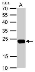
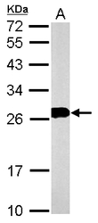
- PGP9.5 antibody detects PGP9.5 protein by western blot analysis.A. 30 ?g PC-12 whole cell lysate/extract12% SDS-PAGEPGP9.5 antibody (GTX109637) dilution: 1:10000The HRP-conjugated anti-rabbit IgG antibody (GTX213110-01) was used to detect the primary antibody.
- Submitted by
- GeneTex (provider)
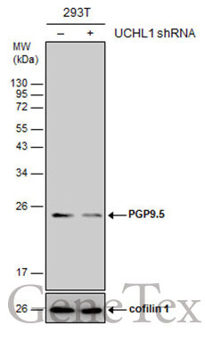
- Main image
- Experimental details
- Non-transfected (¡V) and transfected (+) 293T whole cell extracts (30 ?g) were separated by 12% SDS-PAGE, and the membrane was blotted with PGP9.5 antibody (GTX109637) diluted at 1:2000. The HRP-conjugated anti-rabbit IgG antibody (GTX213110-01) was used to detect the primary antibody.
Supportive validation
- Submitted by
- GeneTex (provider)
- Main image
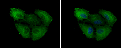
- Experimental details
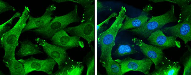
- PGP9.5 antibody detects PGP9.5 protein at cytoplasm by immunofluorescent analysis.Sample: A549 cells were fixed in 4% paraformaldehyde at RT for 15 min.Green: PGP9.5 protein stained by PGP9.5 antibody (GTX109637) diluted at 1:200.Blue: Hoechst 33342 staining.
- Submitted by
- GeneTex (provider)
- Main image

- Experimental details
- PGP9.5 antibody detects PGP9.5 protein at cytoplasm by immunofluorescent analysis.Sample: DIV9 rat E18 primary cortical neurons were fixed in 4% paraformaldehyde at RT for 15 min.Green: PGP9.5 protein stained by PGP9.5 antibody (GTX109637) diluted at 1:500.Red: beta Tubulin 3/ Tuj1, stained by beta Tubulin 3/ Tuj1 antibody [GT11710] (GTX631836) diluted at 1:500.Blue: Fluoroshield with DAPI (GTX30920).

- Submitted by
- GeneTex (provider)
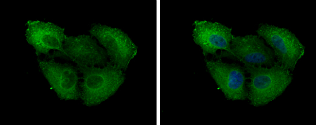
- Main image
- Experimental details
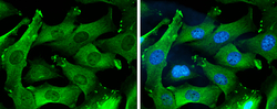
- PGP9.5 antibody detects PGP9.5 protein at cytoplasm by immunofluorescent analysis.Sample: SK-N-SH cells were fixed in 4% paraformaldehyde at RT for 15 min.Green: PGP9.5 protein stained by PGP9.5 antibody (GTX109637) diluted at 1:1000.Blue: Hoechst 33342 staining.
Supportive validation
- Submitted by
- GeneTex (provider)
- Main image
- Experimental details
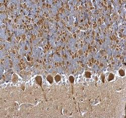
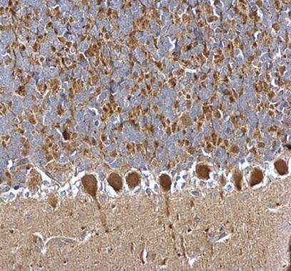
- PGP9.5 antibody detects PGP9.5 protein at cytosol on rat hind brain by immunohistochemical analysis. Sample: Paraffin-embedded rat hind brain. PGP9.5 antibody (GTX109637) dilution: 1:500.
- Submitted by
- GeneTex (provider)
- Main image
- Experimental details
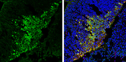
- PGP9.5 antibody detects PGP9.5 protein expression by immunohistochemical analysis.Sample: Frozen sectioned E13.5 Rat brain. Green: PGP9.5 protein stained by PGP9.5 antibody (GTX109637) diluted at 1:250.Red: beta Tubulin 3/ TUJ1, a mature neuron marker, stained by beta Tubulin 3/ TUJ1 antibody [GT11710] (GTX631836) diluted at 1:500.Blue: Fluoroshield with DAPI (GTX30920).

- Submitted by
- GeneTex (provider)
- Main image

- Experimental details
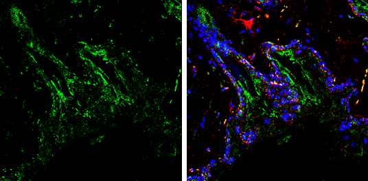
- PGP9.5 antibody detects PGP9.5 protein by immunohistochemical analysis. Samples: Paraffin-embedded mouse skin.Green: PGP9.5 protein stained by PGP9.5 antibody (GTX109637) diluted at 1:250.Red: beta Tubulin 3/ Tuj1, a marker, stained by beta Tubulin 3/ Tuj1 antibody [GT1338] (GTX631831) diluted at 1:500.Blue: Fluoroshield with DAPI (GTX30920).
- Submitted by
- GeneTex (provider)
- Main image
- Experimental details
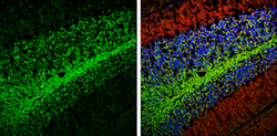
- PGP9.5 antibody detects PGP9.5 protein expression by immunohistochemical analysis.Sample: Frozen-sectioned adult mouse cerebellum. Green: PGP9.5 protein stained by PGP9.5 antibody (GTX109637) diluted at 1:250.Red: beta Tubulin 3/ TUJ1, stained by beta Tubulin 3/ TUJ1 antibody [GT11710] (GTX631836) diluted at 1:500.Blue: Fluoroshield with DAPI (GTX30920).
- Submitted by
- GeneTex (provider)
- Main image

- Experimental details
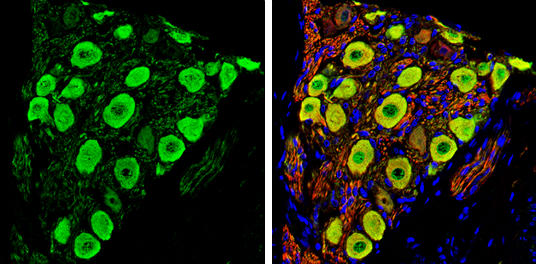
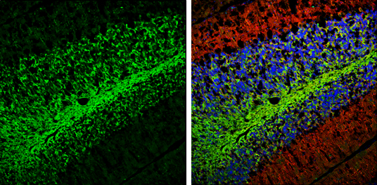
- PGP9.5 antibody detects PGP9.5 protein by immunochemical analysis.Samples: Paraffin-embedded rat colon.Green: PGP9.5 protein stained by PGP9.5 antibody (GTX109637) diluted at 1:250.Red: beta Tubulin 3/ Tuj1, stained by beta Tubulin 3/ Tuj1 antibody [GT1338] (GTX631831) diluted at 1:500.Blue: Fluoroshield with DAPI (GTX30920).