Explore
 Validate
 Learn
Learn Western blot
 Immunocytochemistry
Antibody data
- Antibody Data
- Antigen structure
- References [0]
- Comments [0]
- Validations
- Western blot [6]
- Immunocytochemistry [1]
- Immunohistochemistry [4]
Submit
Validation data
Reference
Comment
Report error
- Product number
- GTX109646 - Provider product page

- Provider
- GeneTex
- Proper citation
- GeneTex Cat#GTX109646, RRID:AB_1952495
- Product name
- PGP9.5 antibody
- Antibody type
- Polyclonal
- Reactivity
- Human, Mouse, Rat
- Host
- Rabbit
No comments: Submit comment
Enhanced validation
Supportive validation
- Submitted by
- GeneTex (provider)
- Enhanced method
- Genetic validation
- Main image

- Experimental details
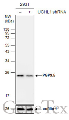
- Non-transfected (¡V) and transfected (+) 293T whole cell extracts (30 ?g) were separated by 12% SDS-PAGE, and the membrane was blotted with PGP9.5 antibody (GTX109646) diluted at 1:3000.

Supportive validation
- Submitted by
- GeneTex (provider)
- Main image

- Experimental details
- Sample (30 ug of whole cell lysate) A: A549 B: H1299 12% SDS PAGE GTX109646 diluted at 1:5000

- Submitted by
- GeneTex (provider)
- Main image

- Experimental details
- PGP9.5 antibody detects UCHL1 protein by Western blot analysis.A. 50 ?g mouse brain lysate/extract12 % SDS-PAGEPGP9.5 antibody (GTX109646) dilution: 1:5000

- Submitted by
- GeneTex (provider)
- Main image

- Experimental details
- PGP9.5 antibody detects UCHL1 protein by Western blot analysis.A. 50 µg rat brain lysate/extract12 % SDS-PAGEPGP9.5 antibody (GTX109646) dilution: 1:10000
- Validation comment
- WB

- Submitted by
- GeneTex (provider)
- Main image

- Experimental details
- Rat tissue extract (50 ?g) was separated by 12% SDS-PAGE, and the membrane was blotted with PGP9.5 antibody (GTX109646) diluted at 1:10000.

- Submitted by
- GeneTex (provider)
- Main image

- Experimental details
- Non-transfected (¡V) and transfected (+) 293T whole cell extracts (30 ?g) were separated by 12% SDS-PAGE, and the membrane was blotted with PGP9.5 antibody (GTX109646) diluted at 1:3000.
Supportive validation
- Submitted by
- GeneTex (provider)
- Main image

- Experimental details
- PGP9.5 antibody detects PGP9.5 protein at cytoplasm by immunofluorescent analysis.Sample: DIV9 rat E18 primary cortical neurons were fixed in 4% paraformaldehyde at RT for 15 min.Green: PGP9.5 protein stained by PGP9.5 antibody (GTX109646) diluted at 1:500.Red: beta Tubulin 3/ Tuj1, stained by beta Tubulin 3/ Tuj1 antibody [GT11710] (GTX631836) diluted at 1:500.Blue: Fluoroshield with DAPI (GTX30920).

Supportive validation
- Submitted by
- GeneTex (provider)
- Main image

- Experimental details
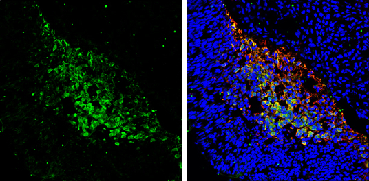
- Immunohistochemical analysis of paraffin-embedded SEROUS OVCA xenograft, using PGP9.5(GTX109646) antibody at 1:300 dilution.

- Submitted by
- GeneTex (provider)
- Main image

- Experimental details
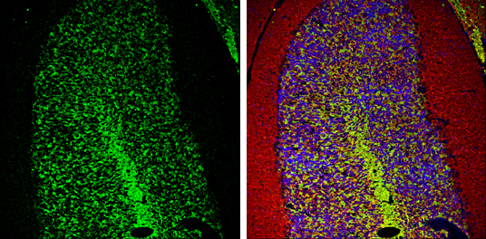
- PGP9.5 antibody detects PGP9.5 protein expression by immunohistochemical analysis.Sample: Frozen sectioned E13.5 Rat brain. Green: PGP9.5 protein stained by PGP9.5 antibody (GTX109646) diluted at 1:250.Red: beta Tubulin 3/ TUJ1, a mature neuron marker, stained by beta Tubulin 3/ TUJ1 antibody [GT11710] (GTX631836) diluted at 1:500.Blue: Fluoroshield with DAPI (GTX30920).
- Submitted by
- GeneTex (provider)
- Main image

- Experimental details
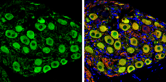
- PGP9.5 antibody detects PGP9.5 protein expression by immunohistochemical analysis.Sample: Frozen-sectioned adult mouse cerebellum. Green: PGP9.5 protein stained by PGP9.5 antibody (GTX109646) diluted at 1:250.Red: beta Tubulin 3/ TUJ1, stained by beta Tubulin 3/ TUJ1 antibody [GT11710] (GTX631836) diluted at 1:500.Blue: Fluoroshield with DAPI (GTX30920).
- Submitted by
- GeneTex (provider)
- Main image
- Experimental details
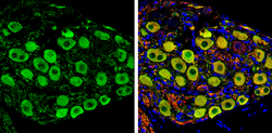
- PGP9.5 antibody detects PGP9.5 protein by immunochemical analysis.Samples: Paraffin-embedded rat colon.Green: PGP9.5 protein stained by PGP9.5 antibody (GTX109646) diluted at 1:250.Red: beta Tubulin 3/ Tuj1, stained by beta Tubulin 3/ Tuj1 antibody [GT1338] (GTX631831) diluted at 1:500.Blue: Fluoroshield with DAPI (GTX30920).