 Explore
 Validate
 Learn
Learn Western blot
 Immunocytochemistry
Antibody data
- Antibody Data
- Antigen structure
- References [2]
- Comments [0]
- Validations
- Western blot [2]
- Immunohistochemistry [1]
- Flow cytometry [1]
Submit
Validation data
Reference
Comment
Report error
- Product number
- NB300-676 - Provider product page

- Provider
- Novus Biologicals
- Proper citation
- Novus Cat#NB300-676, RRID:AB_2138454
- Product name
- Rabbit Polyclonal UCH-L1/PGP9.5 Antibody
- Antibody type
- Polyclonal
- Description
- Immunogen affinity purified.
- Reactivity
- Human, Mouse
- Host
- Rabbit
- Isotype
- IgG
- Vial size
- 0.1 ml
- Concentration
- 1.0 mg/ml
- Storage
- Store at 4C short term. Aliquot and store at -20C long term. Avoid freeze-thaw cycles.
Submitted references Increased levels of UCHL1 are a compensatory response to disrupted ubiquitin homeostasis in spinal muscular atrophy and do not represent a viable therapeutic target.
Increased BrdU incorporation reflecting DNA repair, neuronal de-differentiation or possible neurogenesis in the adult cochlear nucleus following bilateral cochlear lesions in the rat.
Powis RA, Mutsaers CA, Wishart TM, Hunter G, Wirth B, Gillingwater TH
Neuropathology and applied neurobiology 2014 Dec;40(7):873-87
Neuropathology and applied neurobiology 2014 Dec;40(7):873-87
Increased BrdU incorporation reflecting DNA repair, neuronal de-differentiation or possible neurogenesis in the adult cochlear nucleus following bilateral cochlear lesions in the rat.
Zheng Y, Begum S, Zhang C, Fleming K, Masumura C, Zhang M, Smith P, Darlington C
Experimental brain research 2011 May;210(3-4):477-87
Experimental brain research 2011 May;210(3-4):477-87
No comments: Submit comment
Supportive validation
- Submitted by
- Novus Biologicals (provider)
- Main image

- Experimental details
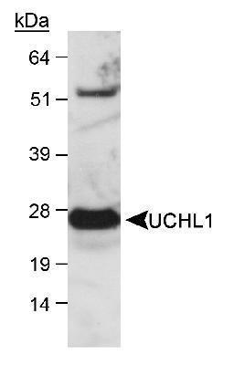
- Western Blot: UCH-L1/PGP9.5 Antibody [NB300-676] - Analysis of UCHL1 in mouse brain lysate ECL detection, 60 seconds.
- Submitted by
- Novus Biologicals (provider)
- Main image
- Experimental details
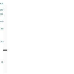
- Simple Western: UCH-L1/PGP9.5 Antibody [NB300-676] - Image shows a specific band for PGP9.5/UCHL-1 in 0.05 mg/mL of Human Brain lysate. This experiment was performed under reducing conditions using the 12-230 kDa separation system.

Supportive validation
- Submitted by
- Novus Biologicals (provider)
- Main image
- Experimental details
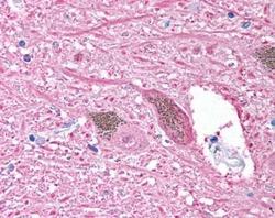
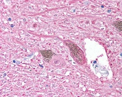
- Immunohistochemistry-Paraffin: UCH-L1/PGP9.5 Antibody [NB300-676] - Staining of neurons and cell processes at 2.5ug/mL. Human brain, Substantia Nigra, Pigmented Neurons, 4X.
Supportive validation
- Submitted by
- Novus Biologicals (provider)
- Main image
- Experimental details
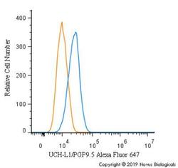
- Flow Cytometry: UCH-L1/PGP9.5 Antibody [NB300-676] - An intracellular stain was performed on U-87 cells with UCH-L1/PGP9.5 Antibody NB300-676AF647 (blue) and a matched isotype control (orange). Cells were fixed with 4% PFA and then permeabilized with 0.1% saponin. Cells were incubated in an antibody dilution of 5 ug/mL for 30 minutes at room temperature. Both antibodies were conjugated to Alexa Fluor 647.
