Explore
 Validate
 Learn
Learn Western blot
Antibody data
- Antibody Data
- Antigen structure
- References [0]
- Comments [0]
- Validations
- Western blot [5]
- Immunocytochemistry [1]
- Immunohistochemistry [1]
Submit
Validation data
Reference
Comment
Report error
- Product number
- PA5-22272 - Provider product page

- Provider
- Invitrogen Antibodies
- Product name
- Aspartoacylase Polyclonal Antibody
- Antibody type
- Polyclonal
- Antigen
- Recombinant protein fragment
- Description
- Recommended positive controls: Molt-4, Raji, U87-MG, SK-N-SH, IMR32, SK-N-AS. Predicted reactivity: Mouse (87%), Rat (84%), Pig (95%), Rhesus Monkey (97%), Bovine (93%). Store product as a concentrated solution. Centrifuge briefly prior to opening the vial.
- Reactivity
- Human, Mouse
- Host
- Rabbit
- Isotype
- IgG
- Vial size
- 100 µL
- Concentration
- 1.33 mg/mL
- Storage
- Store at 4°C short term. For long term storage, store at -20°C, avoiding freeze/thaw cycles.
No comments: Submit comment
Supportive validation
- Submitted by
- Invitrogen Antibodies (provider)
- Main image

- Experimental details
- Western blot analysis of Aspartoacylase using Various whole cell extracts (30 µg). Samples were loaded onto a 10% SDS-PAGE gel and probed with an Aspartoacylase polyclonal antibody (Product # PA5-22272) at a dilution of 1:1000.

- Submitted by
- Invitrogen Antibodies (provider)
- Main image

- Experimental details
- Western blot analysis of Aspartoacylase using 30 µg of A) MOLT4 and B) Raji lysate. Samples were loaded onto a 10% SDS-PAGE gel and probed with an Aspartoacylase polyclonal antibody (Product # PA5-22272) at a dilution of 1:1000.

- Submitted by
- Invitrogen Antibodies (provider)
- Main image
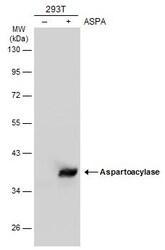
- Experimental details
- Western Blot analysis of Aspartoacylase was performed by separating 30 µg of non-transfected (–) and transfected (+) 293T whole cell extracts by 10% SDS-PAGE. Proteins were transferred to a membrane and probed with a Aspartoacylase Polyclonal Antibody (Product # PA5-22272) at a dilution of 1:10000. The HRP-conjugated anti-rabbit IgG antibody was used to detect the primary antibody.

- Submitted by
- Invitrogen Antibodies (provider)
- Main image

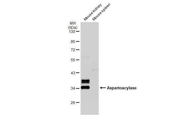
- Experimental details
- Western Blot using Aspartoacylase Polyclonal Antibody (Product # PA5-22272). Various tissue extracts (50 µg) were separated by 10% SDS-PAGE, and the membrane was blotted with Aspartoacylase Polyclonal Antibody (Product # PA5-22272) diluted at 1:1,000. The HRP-conjugated anti-rabbit IgG antibody was used to detect the primary antibody.
- Submitted by
- Invitrogen Antibodies (provider)
- Main image

- Experimental details
- Western Blot using Aspartoacylase Polyclonal Antibody (Product # PA5-22272). Various whole cell extracts (30 µg) were separated by 12% SDS-PAGE, and the membrane was blotted with Aspartoacylase Polyclonal Antibody (Product # PA5-22272) diluted at 1:1,000. The HRP-conjugated anti-rabbit IgG antibody was used to detect the primary antibody.

Supportive validation
- Submitted by
- Invitrogen Antibodies (provider)
- Main image
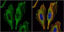
- Experimental details
- Immunocytochemistry-Immunofluorescence analysis of Aspartoacylase was performed in HeLa cells fixed in 4% paraformaldehyde at RT for 15 min. Green: Aspartoacylase Polyclonal Antibody (Product # PA5-22272) diluted at 1:500. Red: alpha Tubulin, a cytoskeleton marker. Blue: Hoechst 33342 staining.

Supportive validation
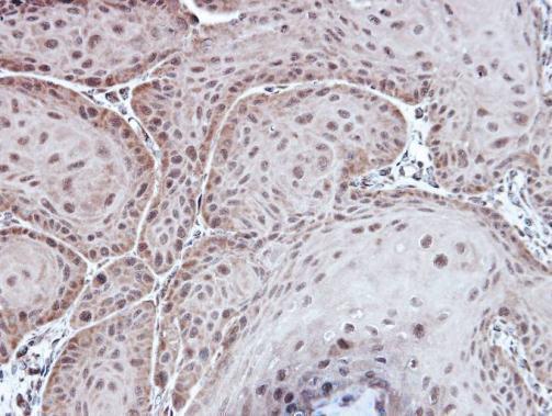
- Submitted by
- Invitrogen Antibodies (provider)
- Main image
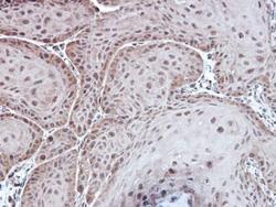
- Experimental details
- Immunohistochemical analysis of paraffin-embedded Cal27 xenograft, using Aspartoacylase (Product # PA5-22272) antibody at 1:100 dilution. Antigen Retrieval: EDTA based buffer, pH 8.0, 15 min.