Explore
 Validate
 Learn
LearnNBP1-83002
antibody from Novus Biologicals
Targeting: RAP1GAP
KIAA0474, RAP1GA1, RAP1GAP1, RAP1GAPII
 Immunocytochemistry
 Immunohistochemistry
Antibody data
- Antibody Data
- Antigen structure
- References [1]
- Comments [0]
- Validations
- Immunohistochemistry [9]
Submit
Validation data
Reference
Comment
Report error
- Product number
- NBP1-83002 - Provider product page

- Provider
- Novus Biologicals
- Proper citation
- Novus Cat#NBP1-83002, RRID:AB_11005719
- Product name
- Rabbit Polyclonal RAP1GAP Antibody
- Antibody type
- Polyclonal
- Description
- Immunogen affinity purified. Specificity of human, mouse RAP1GAP antibody verified on a Protein Array containing target protein plus 383 other non-specific proteins.
- Reactivity
- Human, Mouse
- Host
- Rabbit
- Isotype
- IgG
- Vial size
- 0.1 ml
- Storage
- Store at 4C short term. Aliquot and store at -20C long term. Avoid freeze-thaw cycles.
Submitted references Tissue profiling of the mammalian central nervous system using human antibody-based proteomics.
Mulder J, Björling E, Jonasson K, Wernérus H, Hober S, Hökfelt T, Uhlén M
Molecular & cellular proteomics : MCP 2009 Jul;8(7):1612-22
Molecular & cellular proteomics : MCP 2009 Jul;8(7):1612-22
No comments: Submit comment
Supportive validation
- Submitted by
- Novus Biologicals (provider)
- Main image
- Experimental details
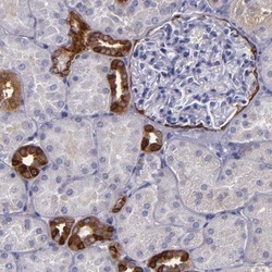
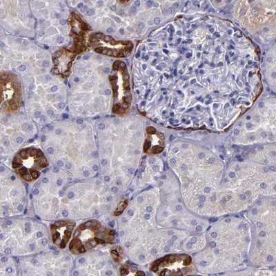
- Immunohistochemistry-Paraffin: RAP1GAP Antibody [NBP1-83002] - Staining of human kidney shows strong positivity in distal tubules and Bowman's capsule.
- Submitted by
- Novus Biologicals (provider)
- Main image

- Experimental details
- Immunohistochemistry-Paraffin: RAP1GAP Antibody [NBP1-83002] - Staining of human cerebellum shows strong cytoplasmic immunoreactivity in Purkinje cells and neurons in the molecular layer.
- Submitted by
- Novus Biologicals (provider)
- Main image
- Experimental details
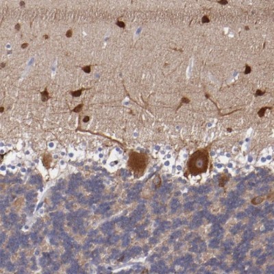
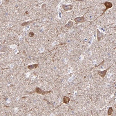
- Immunohistochemistry-Paraffin: RAP1GAP Antibody [NBP1-83002] - Staining of human cerebral cortex shows cytoplasmic immunoreactivity in neuronal cell bodies.
- Submitted by
- Novus Biologicals (provider)
- Main image

- Experimental details
- Immunohistochemistry-Paraffin: RAP1GAP Antibody [NBP1-83002] - Staining of human hippocampus shows cytoplasmic positivity in neuronal cells.
- Submitted by
- Novus Biologicals (provider)
- Main image
- Experimental details
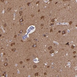
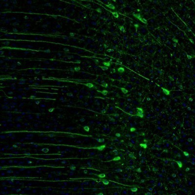
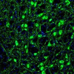
- Immunohistochemistry: RAP1GAP Antibody [NBP1-83002] - Staining of mouse midbrain shows strong immunoreactivity in locus coeruleus neurons.

- Submitted by
- Novus Biologicals (provider)
- Main image
- Experimental details
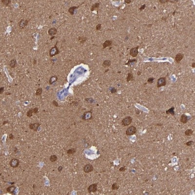
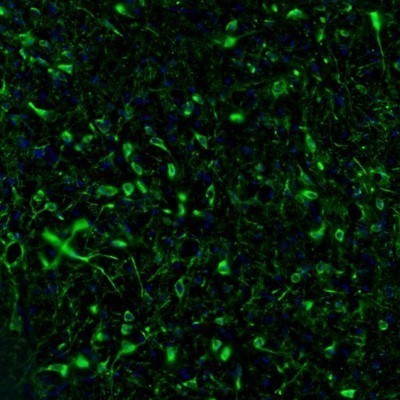
- Immunohistochemistry: RAP1GAP Antibody [NBP1-83002] - Staining of mouse cingulate cortex shows strong cytoplasmic positivity in neuronal cell bodies and proximal dendrites.
- Submitted by
- Novus Biologicals (provider)
- Main image
- Experimental details
- Immunohistochemistry: RAP1GAP Antibody [NBP1-83002] - Staining of mouse hypothalamus shows positivity in neuronal cell bodies and processes in the lateral preoptic area.
- Submitted by
- Novus Biologicals (provider)
- Main image
- Experimental details
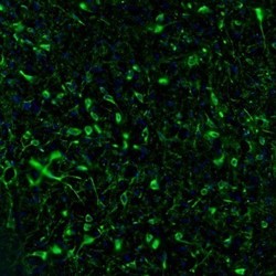
- Immunohistochemistry: RAP1GAP Antibody [NBP1-83002] - Staining of mouse globus pallidus shows strong cytoplasmic immunoreactivity in neurons.

- Submitted by
- Novus Biologicals (provider)
- Main image
- Experimental details
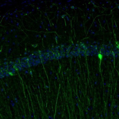
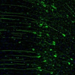
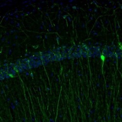
- Immunohistochemistry: RAP1GAP Antibody [NBP1-83002] - Staining of mouse hippocampus shows cytoplasmic positivity in a subset of interneurons in the CA1 area.