Explore
 Validate
 Learn
Learn Western blot
Antibody data
- Antibody Data
- Antigen structure
- References [3]
- Comments [0]
- Validations
- Western blot [2]
Submit
Validation data
Reference
Comment
Report error
- Product number
- NB100-644 - Provider product page

- Provider
- Novus Biologicals
- Proper citation
- Novus Cat#NB100-644, RRID:AB_10001270
- Product name
- Rabbit Polyclonal PINK1 Antibody
- Antibody type
- Polyclonal
- Description
- Immunogen affinity purified. Reacts with residues residues 258-274 (YRKSKRGPKQLAPHPNI) of human PINK1 and will only bind to isoform 1.
- Reactivity
- Human
- Host
- Rabbit
- Isotype
- IgG
- Vial size
- 0.1 mg
- Concentration
- 1 mg/ml
- Storage
- Store at 4C short term. Aliquot and store at -20C long term. Avoid freeze-thaw cycles.
Submitted references Parkin, PINK1, and DJ-1 form a ubiquitin E3 ligase complex promoting unfolded protein degradation.
The kinase domain of mitochondrial PINK1 faces the cytoplasm.
C-terminal truncation and Parkinson's disease-associated mutations down-regulate the protein serine/threonine kinase activity of PTEN-induced kinase-1.
Xiong H, Wang D, Chen L, Choo YS, Ma H, Tang C, Xia K, Jiang W, Ronai Z, Zhuang X, Zhang Z
The Journal of clinical investigation 2009 Mar;119(3):650-60
The Journal of clinical investigation 2009 Mar;119(3):650-60
The kinase domain of mitochondrial PINK1 faces the cytoplasm.
Zhou C, Huang Y, Shao Y, May J, Prou D, Perier C, Dauer W, Schon EA, Przedborski S
Proceedings of the National Academy of Sciences of the United States of America 2008 Aug 19;105(33):12022-7
Proceedings of the National Academy of Sciences of the United States of America 2008 Aug 19;105(33):12022-7
C-terminal truncation and Parkinson's disease-associated mutations down-regulate the protein serine/threonine kinase activity of PTEN-induced kinase-1.
Sim CH, Lio DS, Mok SS, Masters CL, Hill AF, Culvenor JG, Cheng HC
Human molecular genetics 2006 Nov 1;15(21):3251-62
Human molecular genetics 2006 Nov 1;15(21):3251-62
No comments: Submit comment
Supportive validation
- Submitted by
- Novus Biologicals (provider)
- Main image
- Experimental details
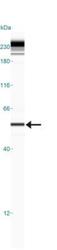
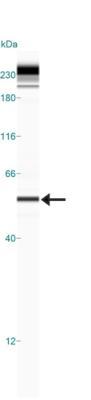
- Simple Western: PINK1 Antibody [NB100-644] - Simple Western lane view shows a specific band for PINK1 at a dilution of 1:50 in 1.0 mg/ml of HeLa lysate. Molecular weight ~56 kDa. This experiment was performed under reducing conditions using the 12-230 kDa separation system.
- Submitted by
- Novus Biologicals (provider)
- Main image
- Experimental details
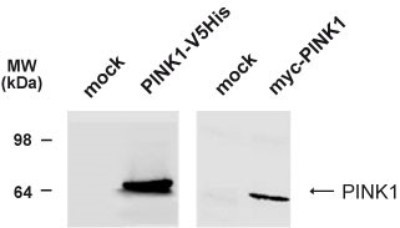
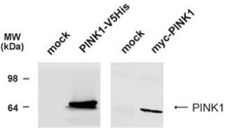
- Western Blot: PINK1 Antibody [NB100-644] - Analysis of C-terminally V5His-tagged human PINK1 or N-teminally myc-tagged human PINK1 expressed in HEK293T cells using NB100-644 at 1:1000 dilution. Observed molecular weight ~64 kDa.