Explore
 Validate
 Learn
Learn Western blot
Antibody data
- Antibody Data
- Antigen structure
- References [3]
- Comments [0]
- Validations
- Western blot [4]
- Immunocytochemistry [1]
- Immunohistochemistry [4]
- Other assay [1]
Submit
Validation data
Reference
Comment
Report error
- Product number
- PA5-54909 - Provider product page

- Provider
- Invitrogen Antibodies
- Product name
- Anti-TFE3 Polyclonal Antibody
- Antibody type
- Polyclonal
- Antigen
- Recombinant full-length protein
- Description
- Immunogen sequence: SKDLESRQRS LEQANRSLQL RIQELELQAQ IHGLPVPPTP GLLSLATTSA SDSLKPEQLD IEEEGRPGAA TFHVGGGPAQ NAPHQQPPAP PSDALLDLHF PSDHLGDLGD PFHLGLEDIL MEEEEGVVGG LSGGALSPLR AAS Highest antigen sequence identity to the following orthologs: Mouse - 92%, Rat - 92%.
- Reactivity
- Human, Mouse, Rat
- Host
- Rabbit
- Isotype
- IgG
- Vial size
- 100 µL
- Concentration
- 0.3 mg/mL
- Storage
- Store at 4°C short term. For long term storage, store at -20°C, avoiding freeze/thaw cycles.
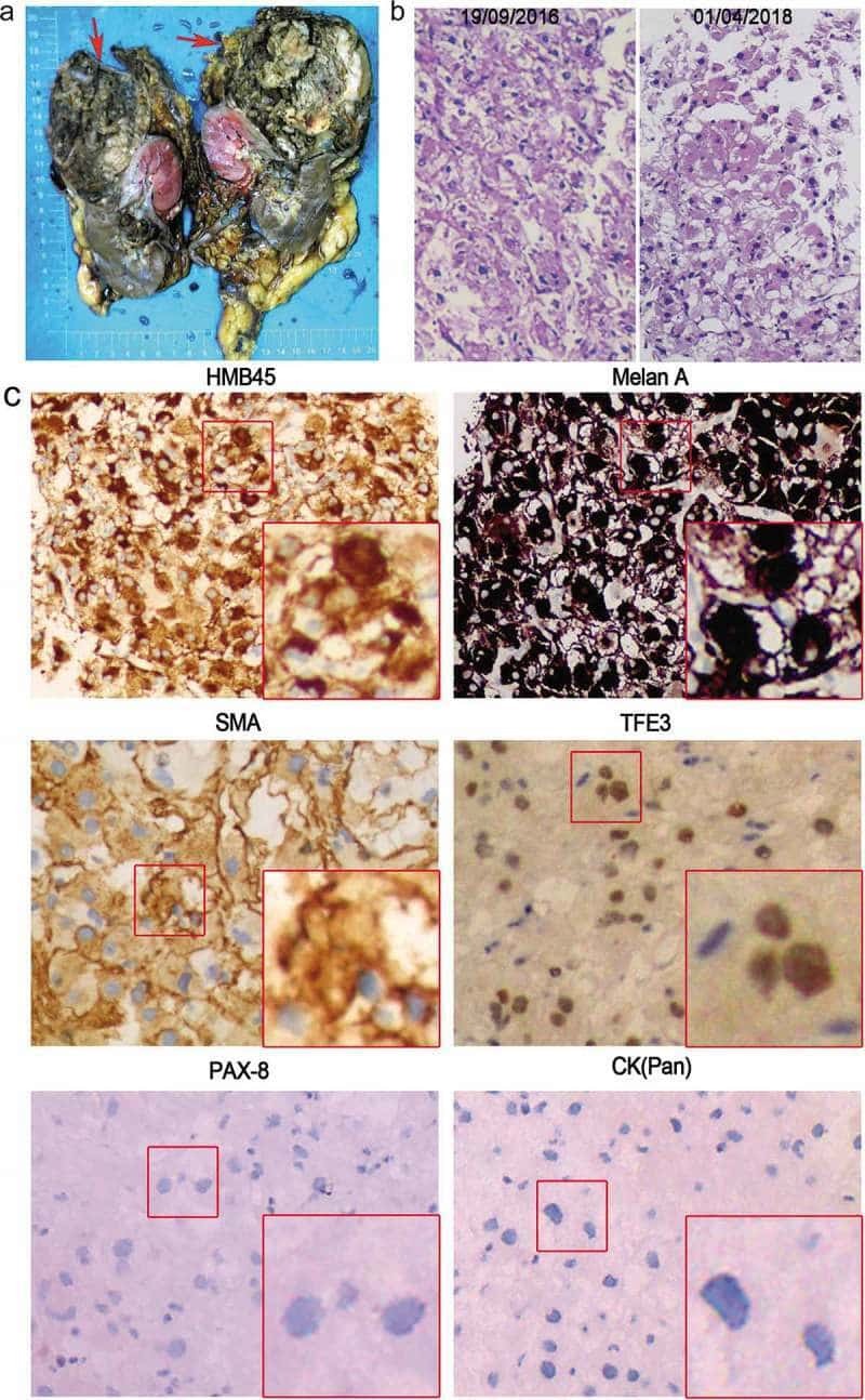
Submitted references Two novel TSC2 mutations in renal epithelioid angiomyolipoma sensitive to everolimus.
Autophagy induction by thiostrepton improves the efficacy of immunogenic chemotherapy.
Towards Age-Related Anti-Inflammatory Therapy: Klotho Suppresses Activation of ER and Golgi Stress Response in Senescent Monocytes.
Wang T, Xie S, Luo R, Shi L, Bai P, Wang X, Wan R, Deng J, Wu Z, Li W, Xiao W, Wang Y, Chen B, Zhang K, Xing J
Cancer biology & therapy 2020;21(1):4-11
Cancer biology & therapy 2020;21(1):4-11
Autophagy induction by thiostrepton improves the efficacy of immunogenic chemotherapy.
Wang Y, Xie W, Humeau J, Chen G, Liu P, Pol J, Zhang Z, Kepp O, Kroemer G
Journal for immunotherapy of cancer 2020 Mar;8(1)
Journal for immunotherapy of cancer 2020 Mar;8(1)
Towards Age-Related Anti-Inflammatory Therapy: Klotho Suppresses Activation of ER and Golgi Stress Response in Senescent Monocytes.
Mytych J, Sołek P, Będzińska A, Rusinek K, Warzybok A, Tabęcka-Łonczyńska A, Koziorowski M
Cells 2020 Jan 21;9(2)
Cells 2020 Jan 21;9(2)
No comments: Submit comment
Supportive validation
- Submitted by
- Invitrogen Antibodies (provider)
- Main image

- Experimental details
- Western blot analysis of TFE3 in U-87MG ATCC cells transfected with control siRNA, target specific siRNA probe #1 and #2, using a TFE3 Polyclonal Antibody (Product # PA5-54909). Remaining relative intensity is presented. Loading control: Anti-GAPDH.

- Submitted by
- Invitrogen Antibodies (provider)
- Main image

- Experimental details
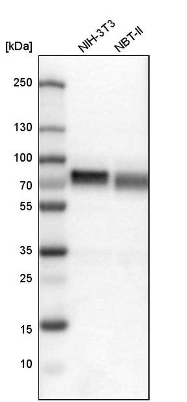
- Western blot analysis of TFE3 in mouse cell line NIH-3T3 and rat cell line NBT-II using a TFE3 Polyclonal Antibody (Product # PA5-54909).
- Submitted by
- Invitrogen Antibodies (provider)
- Main image

- Experimental details
- Knockdown of TFE3 was achieved by transfecting HeLa cells with TFE3 specific siRNAs (Silencer® select Product # s14030, s14031). Western blot analysis (Fig. a) was performed using whole cell extracts from the TFE3 knockdown cells (Lane 3), non-specific scrambled siRNA transfected cells (Lane 2) and untransfected cells (Lane 1). The blot was probed using TFE3 Polyclonal Antibody (Product # PA5-54909, 0.4µg/ml) and Goat Anti-Rabbit IgG Secondary Antibody, HRP conjugate (Product # A27036, 1:4000 dilution). Densitometric analysis of this western blot for the 61 kDa band is shown in histogram (Fig. b). Loss of signal upon siRNA mediated knock down confirms that antibody is specific to TFE3.

- Submitted by
- Invitrogen Antibodies (provider)
- Main image
- Experimental details
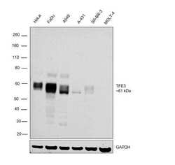
- Western blot was performed using Anti-TFE3 Polyclonal Antibody (Product # PA5-54909) and a ~61 kDa band corresponding to TFE3 was observed across all the cell lines tested except for MOLT-4. Whole cell extracts (30 µg) of HeLa (Lane1), FaDu (Lane 2), A549 (Lane 3), A-431 (Lane 4), SK-BR-3 (Lane 5) and MOLT-4 (Lane 6)were electrophoresed using NuPAGE™ 10% Bis-Tris Protein Gel (Product # NP0302BOX). Resolved proteins were then transferred onto a nitrocellulose membrane (Product # IB23001) by iBlot® 2 Dry Blotting System (Product # IB21001). The blot was probed with the primary antibody (0.4µg/ml) and detected by chemiluminescence Goat Anti-Rabbit IgG Secondary Antibody, HRP conjugate (Product # A27036, 1:4000 dilution) using the iBright FL 1000 (Product # A32752). Chemiluminescent detection was performed using Novex® ECL Chemiluminescent Substrate Reagent Kit (Product # WP20005).

Supportive validation
- Submitted by
- Invitrogen Antibodies (provider)
- Main image

- Experimental details
- Immunofluorescent staining of TFE3 in human cell line A-431 shows positivity in cytoplasm & nucleus but excluded from the nucleoli. Samples were probed using a TFE3 Polyclonal Antibody (Product # PA5-54909).

Supportive validation
- Submitted by
- Invitrogen Antibodies (provider)
- Main image

- Experimental details
- Immunohistochemical staining of TFE3 in human placenta using a TFE3 Polyclonal Antibody (Product # PA5-54909) shows moderate to strong nuclear positivity in trophoblastic cells.

- Submitted by
- Invitrogen Antibodies (provider)
- Main image
- Experimental details
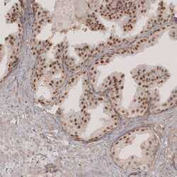
- Immunohistochemical staining of TFE3 in human prostate using a TFE3 Polyclonal Antibody (Product # PA5-54909) shows moderate to strong nuclear positivity in glandular cells.

- Submitted by
- Invitrogen Antibodies (provider)
- Main image

- Experimental details
- Immunohistochemical staining of TFE3 in human kidney using a TFE3 Polyclonal Antibody (Product # PA5-54909) shows moderate nuclear positivity in renal glomeruli and tubuli cells.

- Submitted by
- Invitrogen Antibodies (provider)
- Main image

- Experimental details
- Immunohistochemical staining of TFE3 in human testis using a TFE3 Polyclonal Antibody (Product # PA5-54909) shows moderate nuclear positivity in a subset of cells in seminiferous ducts.

Supportive validation
- Submitted by
- Invitrogen Antibodies (provider)
- Main image
- Experimental details
- NULL