Explore
 Validate
 Learn
Learn Western blot
 Immunocytochemistry
Antibody data
- Antibody Data
- Antigen structure
- References [2]
- Comments [0]
- Validations
- Immunocytochemistry [1]
- Blocking/Neutralizing [1]
Submit
Validation data
Reference
Comment
Report error
- Product number
- AF1886 - Provider product page

- Provider
- R&D Systems
- Product name
- Equine IL-6 Antibody
- Antibody type
- Polyclonal
- Description
- Immunogen affinity purified. Detects equine IL-6 in direct ELISAs and Western blots. In direct ELISAs and Western blots, approximately 50% cross-reactivity with recombinant human IL-6, recombinant porcine IL-6 and recombinant canine IL-6 is observed and approximately 20% cross-reactivity with recombinant mouse IL-6 and recombinant rat IL-6 is observed.
- Host
- Goat
- Conjugate
- Unconjugated
- Antigen sequence
Q95181- Isotype
- IgG
- Vial size
- 100 ug
- Concentration
- LYOPH
- Storage
- Use a manual defrost freezer and avoid repeated freeze-thaw cycles. 12 months from date of receipt, -20 to -70 °C as supplied. 1 month, 2 to 8 °C under sterile conditions after reconstitution. 6 months, -20 to -70 °C under sterile conditions after reconstitution.
Submitted references Equine mesenchymal stem cells inhibit T cell proliferation through different mechanisms depending on tissue source.
Serum interleukin-6 (IL-6) and IL-10 concentrations in normal and septic neonatal foals.
Carrade Holt DD, Wood JA, Granick JL, Walker NJ, Clark KC, Borjesson DL
Stem cells and development 2014 Jun 1;23(11):1258-65
Stem cells and development 2014 Jun 1;23(11):1258-65
Serum interleukin-6 (IL-6) and IL-10 concentrations in normal and septic neonatal foals.
Burton AB, Wagner B, Erb HN, Ainsworth DM
Veterinary immunology and immunopathology 2009 Dec 15;132(2-4):122-8
Veterinary immunology and immunopathology 2009 Dec 15;132(2-4):122-8
No comments: Submit comment
Supportive validation
- Submitted by
- R&D Systems (provider)
- Main image

- Experimental details
- IL-6 in Equine PBMCs. IL-6 was detected in immersion fixed equine peripheral blood mononuclear cells (PBMCs) using Goat Anti-Equine IL-6 Antigen Affinity-purified Polyclonal Antibody (Catalog # AF1886) at 10 µg/mL for 3 hours at room temperature. Cells were stained using the NorthernLights™ 557-conjugated Anti-Goat IgG Secondary Antibody (red; Catalog # NL001) and counterstained with DAPI (blue). Specific staining was localized to the cell surface. View our protocol for Fluorescent ICC Staining of Non-adherent Cells.

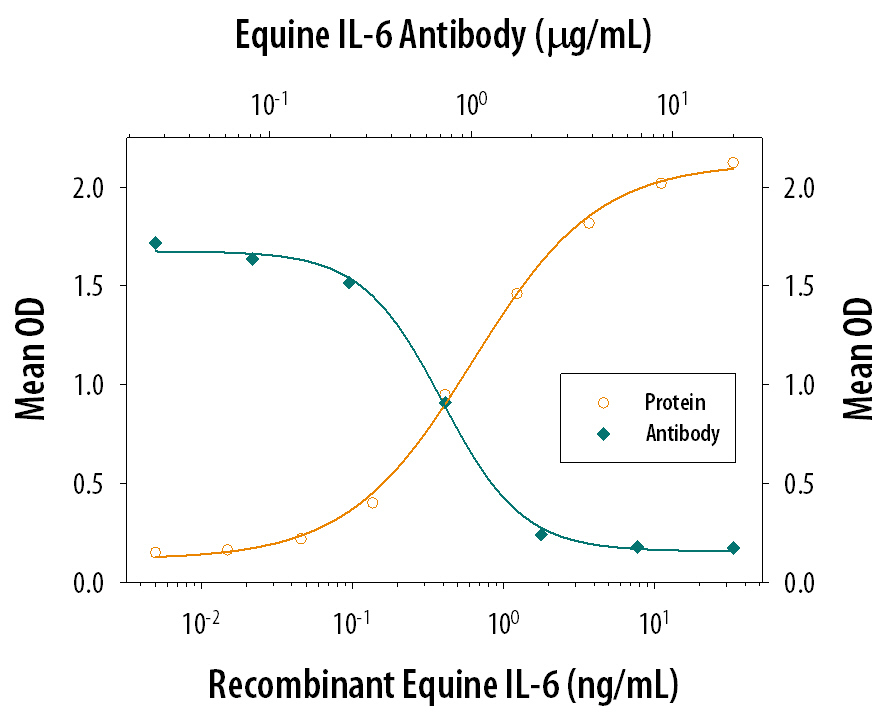
Supportive validation
- Submitted by
- R&D Systems (provider)
- Main image

- Experimental details
- Cell Proliferation Induced by IL-6 and Neutralization by Equine IL-6 Antibody. Recombinant Equine IL-6 (Catalog # 1886-EL) stimulates proliferation in the T1165.85.2.1 mouse plasmacytoma cell line in a dose-dependent manner (orange line). Proliferation elicited by Recombinant Equine IL-6 (4 ng/mL) is neutralized (green line) by increasing concentrations of Goat Anti-Equine IL-6 Antigen Affinity-purified Polyclonal Antibody (Catalog # AF1886). The ND50 is typically 0.5-1.5 µg/mL.