 Explore
 Validate
 Learn
Learn Western blot
Antibody data
- Antibody Data
- Antigen structure
- References [4]
- Comments [0]
- Validations
- Western blot [2]
- Immunohistochemistry [1]
- Flow cytometry [1]
- Other assay [1]
Submit
Validation data
Reference
Comment
Report error
- Product number
- PA1-41062 - Provider product page

- Provider
- Invitrogen Antibodies
- Product name
- CCR1 Polyclonal Antibody
- Antibody type
- Polyclonal
- Antigen
- Synthetic peptide
- Description
- Suggested positive control: A375, antigen standard for CCR1 (transient overexpression lysate).
- Reactivity
- Human, Mouse
- Host
- Rabbit
- Isotype
- IgG
- Vial size
- 100 µg
- Concentration
- 1 mg/mL
- Storage
- Store at 4°C short term. For long term storage, store at -20°C, avoiding freeze/thaw cycles.
Submitted references Met-RANTES preserves the blood-brain barrier through inhibiting CCR1/SRC/Rac1 pathway after intracerebral hemorrhage in mice.
Tumor-associated neutrophils suppress antitumor immunity of NK cells through the PD-L1/PD-1 axis.
CCR1 Activation Promotes Neuroinflammation Through CCR1/TPR1/ERK1/2 Signaling Pathway After Intracerebral Hemorrhage in Mice.
Crosstalk between CCL7 and CCR3 promotes metastasis of colon cancer cells via ERK-JNK signaling pathways.
Yan J, Xu W, Lenahan C, Huang L, Ocak U, Wen J, Li G, He W, Le C, Zhang JH, Mo L, Tang J
Fluids and barriers of the CNS 2022 Jan 21;19(1):7
Fluids and barriers of the CNS 2022 Jan 21;19(1):7
Tumor-associated neutrophils suppress antitumor immunity of NK cells through the PD-L1/PD-1 axis.
Sun R, Xiong Y, Liu H, Gao C, Su L, Weng J, Yuan X, Zhang D, Feng J
Translational oncology 2020 Oct;13(10):100825
Translational oncology 2020 Oct;13(10):100825
CCR1 Activation Promotes Neuroinflammation Through CCR1/TPR1/ERK1/2 Signaling Pathway After Intracerebral Hemorrhage in Mice.
Yan J, Zuo G, Sherchan P, Huang L, Ocak U, Xu W, Travis ZD, Wang W, Zhang JH, Tang J
Neurotherapeutics : the journal of the American Society for Experimental NeuroTherapeutics 2020 Jul;17(3):1170-1183
Neurotherapeutics : the journal of the American Society for Experimental NeuroTherapeutics 2020 Jul;17(3):1170-1183
Crosstalk between CCL7 and CCR3 promotes metastasis of colon cancer cells via ERK-JNK signaling pathways.
Lee YS, Kim SY, Song SJ, Hong HK, Lee Y, Oh BY, Lee WY, Cho YB
Oncotarget 2016 Jun 14;7(24):36842-36853
Oncotarget 2016 Jun 14;7(24):36842-36853
No comments: Submit comment
Supportive validation
- Submitted by
- Invitrogen Antibodies (provider)
- Main image

- Experimental details
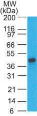
- Western blot analysis of Human heart lysate probed with a CD191/CCR1 polyclonal antibody (Product # PA1-41062) at 2 µg/mL.

- Submitted by
- Invitrogen Antibodies (provider)
- Main image

- Experimental details
- Western blot analysis of Human heart lysate probed with a CD191/CCR1 polyclonal antibody (Product # PA1-41062) at 2 µg/mL.
Supportive validation
- Submitted by
- Invitrogen Antibodies (provider)
- Main image

- Experimental details
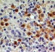
- Immunohistochemical analysis of formalin-fixed, paraffin-embedded normal human spleen tissue stained with a CD191/CCR1 polyclonal antibody (Product # PA1-41062) (5 µg/mL), peroxidase-conjugate and DAB chromogen. Note granulocyte staining.
Supportive validation
- Submitted by
- Invitrogen Antibodies (provider)
- Main image

- Experimental details
- Image 1: Surface staining of mouse primary epidermal cells (B-6 mice, 30wks) with CL 11-PE, CD11c PE-Cy5, and a CD191/CCR1 polyclonal antibody (Product # PA1-41062) with CY5 goat anti-rabbit secondary. Image 2: CD11c+ CL II+ cells (Langerans), stained with rabbit isotype control (shaded histogram) were analyzed for CCR1 at 5 µL (blue histogram) and 10 µL (green histogram).

Supportive validation
- Submitted by
- Invitrogen Antibodies (provider)
- Main image

- Experimental details
- Fig. 2 Temporal expressions of CCR1, CCL5, p-SRC, and Rac1 after ICH in mice. A Representative western blot images and quantitative analyses of time course of CCR1, CCL5, p-SRC, and Rac1 expressions within the ipsilateral hemisphere after ICH. Data were expressed as mean +- SD. * p < 0.05, ** p < 0.01, *** p < 0.001 vs. sham group. n = 6/group. B The four black squares indicated perihematomal regions for microphotograph of the immunofluorescence staining. Double immunofluorescence staining for CCR1 (green) in endothelial cells (vWF, red), astrocytes (GFAP, red), and neurons (NeuN, red) within peri-hematoma area 24 h after ICH. Nuclei were stained with DAPI (blue). Scale bar = 50 mum. n = 2/group
