Explore
 Validate
 Learn
Learn Immunocytochemistry
Antibody data
- Antibody Data
- Antigen structure
- References [5]
- Comments [0]
- Validations
- Immunocytochemistry [1]
Submit
Validation data
Reference
Comment
Report error
- Product number
- 210-401-323S - Provider product page

- Provider
- Rockland Immunochemicals, Inc.
- Proper citation
- Rockland Cat#210-401-323S, RRID:AB_11181154
- Product name
- Anti-Mouse IL-18 (RABBIT) Antibody - 210-401-323S
- Antibody type
- Polyclonal
- Vial size
- 25 µl
Submitted references Epithelial-derived gasdermin D mediates nonlytic IL-1β release during experimental colitis.
A truncating mutation in the autophagy gene UVRAG drives inflammation and tumorigenesis in mice.
Extracellular TDP-43 aggregates target MAPK/MAK/MRK overlapping kinase (MOK) and trigger caspase-3/IL-18 signaling in microglia.
Insulin sensitizers prevent fine particulate matter-induced vascular insulin resistance and changes in endothelial progenitor cell homeostasis.
TRAF3IP2 mediates aldosterone/salt-induced cardiac hypertrophy and fibrosis.
Bulek K, Zhao J, Liao Y, Rana N, Corridoni D, Antanaviciute A, Chen X, Wang H, Qian W, Miller-Little WA, Swaidani S, Tang F, Willard BB, McCrae K, Kang Z, Dubyak GR, Cominelli F, Simmons A, Pizarro TT, Li X
The Journal of clinical investigation 2020 Aug 3;130(8):4218-4234
The Journal of clinical investigation 2020 Aug 3;130(8):4218-4234
A truncating mutation in the autophagy gene UVRAG drives inflammation and tumorigenesis in mice.
Quach C, Song Y, Guo H, Li S, Maazi H, Fung M, Sands N, O'Connell D, Restrepo-Vassalli S, Chai B, Nemecio D, Punj V, Akbari O, Idos GE, Mumenthaler SM, Wu N, Martin SE, Hagiya A, Hicks J, Cui H, Liang C
Nature communications 2019 Dec 12;10(1):5681
Nature communications 2019 Dec 12;10(1):5681
Extracellular TDP-43 aggregates target MAPK/MAK/MRK overlapping kinase (MOK) and trigger caspase-3/IL-18 signaling in microglia.
Leal-Lasarte MM, Franco JM, Labrador-Garrido A, Pozo D, Roodveldt C
FASEB journal : official publication of the Federation of American Societies for Experimental Biology 2017 Jul;31(7):2797-2816
FASEB journal : official publication of the Federation of American Societies for Experimental Biology 2017 Jul;31(7):2797-2816
Insulin sensitizers prevent fine particulate matter-induced vascular insulin resistance and changes in endothelial progenitor cell homeostasis.
Haberzettl P, McCracken JP, Bhatnagar A, Conklin DJ
American journal of physiology. Heart and circulatory physiology 2016 Jun 1;310(11):H1423-38
American journal of physiology. Heart and circulatory physiology 2016 Jun 1;310(11):H1423-38
TRAF3IP2 mediates aldosterone/salt-induced cardiac hypertrophy and fibrosis.
Sakamuri SS, Valente AJ, Siddesha JM, Delafontaine P, Siebenlist U, Gardner JD, Bysani C
Molecular and cellular endocrinology 2016 Jul 5;429:84-92
Molecular and cellular endocrinology 2016 Jul 5;429:84-92
No comments: Submit comment
Supportive validation
- Submitted by
- Rockland Immunochemicals, Inc. (provider)
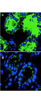
- Main image

- Experimental details
- Immunofluorescence microscopy of IL-18 in mouse colon sections. The transversing portion of the large intestine from DSS-exposed (Panel A) and -unexposed mice (Panel B) was excised, rinsed in PBS, and frozen on isopentane cooled with liquid nitrogen. Frozen sections (5 µm) were cut on a Leica CM 1850 cryostat. The slides were fixed for 10 min in 4% paraformaldehyde, air-dried, and incubated for 20 min in PBS supplemented with 10% normal goat serum. Sections were incubated in a 1:50 dilution of Rockland's rabbit anti-Mouse IL-18 antibody or 1 µg/ml nonimmune rabbit IgG (not shown) as negative control. The antibodies were diluted in PBS containing 1% bovine serum albumin. After an overnight incubation at 4°C, the sections were washed three times with 0.5% bovine serum albumin in PBS. The sections were then incubated with a secondary goat anti-rabbit antibody conjugated to Alexa488 (Molecular Probes) for 60 min at room temperature in the dark. Nuclei were counterstained blue using 1 µg/100 ml bisbenzimide. After staining, sections were washed and examined with the Leica DM RXA confocal laser scanning system and analyzed. Similar staining will occur with other systems.
- Sample type
- IF Microscopy