 Explore
 Validate
 Learn
Learn Western blot
 Immunohistochemistry
Antibody data
- Antibody Data
- Antigen structure
- References [0]
- Comments [0]
- Validations
- Western blot [3]
- ELISA [2]
- Immunocytochemistry [1]
Submit
Validation data
Reference
Comment
Report error
- Product number
- 101-M65A - Provider product page

- Provider
- ReliaTech GmbH
- Product name
- Anti-human PlGF-2
- Antibody type
- Monoclonal
- Description
- antibody Protein-G purified from hybridoma supernatant
- Reactivity
- Human
- Host
- Mouse
- Antigen sequence
LPAVPPQQWALSAGNGSSEVEVVPFQEVWGRSYCR
ALERLVDVVSEYPSEVEHMFSPSCVSLLRCTGCCG
DENLHCVPVETANVTMQLLKIRSGDRPSYVELTFS
QHVRCECRPLREKMKPERRRPKGRGKRRREKQRPT
DCHLCGDAVPRR- Antibody clone number
- (#A6D1)
- Storage
- The lyophilized antibody is stable for at least 2 years at -20°C. After sterile reconstitution the antibody is stable at 2-8°C for up to 6 months. Frozen aliquots are stable for at least 6 months when stored at -20°C. Addition of a carrier protein or 50% glycerol is recommended for frozen aliquots.
- Handling
- Centrifuge vial prior to opening. Reconstitute in sterile water to a concentration of 0.1-1.0 mg/ml.
No comments: Submit comment
Supportive validation
- Submitted by
- ReliaTech GmbH (provider)
- Main image

- Experimental details
- Western Blot analysis with human PlGF1 and PlGF2. Purified recombinant human PlGF1 (red: 500ng, non-red: 1000ng) and PlGF2 (red: 250ng, non-red: 500ng) were loaded in 15% SDS-PAGE under reducing and non-reducing conditions. As primary antibody mouse anti-human PlGF2 #D1 [Cat# 101-M65A] was used in a concentration of 0.5µg/ml. The detection was carried out with a secondary, AP-conjugated anti-mouse Fc antibody (Dianova, 1:1000). NOTE: The anti-human PlGF-2 #D1 antibody [Cat# 101-M65A] recognizes solely the PlGF2 isoform.

- Submitted by
- ReliaTech GmbH (provider)
- Main image

- Experimental details
- Western Blot analysis with PlGF. Purified recombinant human PlGF2 (500 -10 ng/lane), PlGF1 (1000 ng/lane), and mouse and rat PlGF (500ng/lane) all derived from insect cells were loaded in 12.5% SDS-PAGE under reducing conditions. As primary antibody mouse anti-human PlGF2 #D1 [Cat# 101-M65A] was used in a concentration of 2µg/ml. The detection was carried out with a secondary, AP-conjugated anti-mouse Fc antibody. NOTE: There is no cross reaction with human PlGF1, mouse and rat PlGF.

- Submitted by
- ReliaTech GmbH (provider)
- Main image
- Experimental details
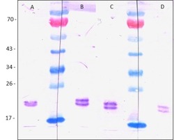
- Western Blot analysis with PlGF. Purified recombinant human PlGF2 (100 ng/lane) derived from insect cells was loaded in 12.5% SDS-PAGE under reducing conditions. As primary antibody mouse anti-human PlGF2 #D1 [Cat# 101-M65A] was used in a concentration of 1µg/ml (A), 0.5µg/ml (B), 0.1µg/ml (C) and 0.05µg/ml (D). The detection was carried out with a secondary, AP-conjugated anti-mouse Fc antibody. NOTE: There is still a signal visible for PlGF2 at an antibody concentration of 50ng/ml.

Supportive validation
- Submitted by
- ReliaTech GmbH (provider)
- Main image
- Experimental details
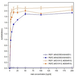
- ELISA with recombinant human PlGF1 and PlGF2 derived from insect cells. Both proteins were coated to a 96-well plate in a concentration of 0.2µg/ml. The mouse anti-human PlGF #331/H12 antibody [Cat# 101-M69] recognizing both isoforms and the mouse anti-human PlGF2 #D1 antibody [Cat# 101-M65A] recognizing specifically PlGF2 were added with increasing concentrations. Detection was carried out with an anti-mouse Biotin- conjugated antibody (Dianova).

- Submitted by
- ReliaTech GmbH (provider)
- Main image

- Experimental details
- ELISA with recombinant human PlGF1 and PlGF2 derived from insect cells. Both proteins were coated to a 96-well plate in increasing amounts. The mouse anti-human PlGF #178/G10 antibody [Cat# 101-M67] recognizing both isoforms and the mouse anti-human PlGF2 #D1 antibody [Cat# 101-M65A] recognizing specifically PlGF2 were added with a concentration of 0.5µg/ml. Detection was carried out with an anti-mouse Biotin antibody (Dianova).

Supportive validation
- Submitted by
- ReliaTech GmbH (provider)
- Main image

- Experimental details
- Immunofluorescence staining of human PlGF1 and PlGF2 in Sf9 cells expressing either PlGF1 or PlGF2. Upper lane: mouse anti-human PlGF2 #D1 [Cat# 101-M65A] recognizing only PlGF2 and lower lane: mouse anti-human PlGF #178/G10 [Cat# 101-M67] recognizing both isoforms of PlGF.
