Explore
 Validate
 Learn
Learn Western blot
Antibody data
- Antibody Data
- Antigen structure
- References [0]
- Comments [0]
- Validations
- Western blot [5]
- Immunocytochemistry [1]
- Immunohistochemistry [5]
Submit
Validation data
Reference
Comment
Report error
- Product number
- HPA026745 - Provider product page

- Provider
- Atlas Antibodies
- Proper citation
- Atlas Antibodies Cat#HPA026745, RRID:AB_1855520
- Product name
- Anti-POLD2
- Antibody type
- Polyclonal
- Reactivity
- Human, Mouse, Rat
- Host
- Rabbit
- Conjugate
- Unconjugated
- Antigen sequence
PRSKYIHPDDELVLEDELQRIKLKGTIDVSKLVTG
TVLAVFGSVRDDGKFLVEDYCFADLAPQKPAPPLD
TDRFVLLVSGLGLGGGGGESLLGTQLLVDVVTGQL
GDEGEQC- Isotype
- IgG
- Vial size
- 100 µl
- Storage
- Store at +4°C for short term storage. Long time storage is recommended at -20°C.
No comments: Submit comment
Supportive validation
- Submitted by
- Atlas Antibodies (provider)
- Enhanced method
- Genetic validation
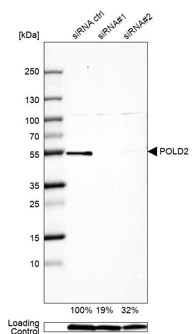
- Main image

- Experimental details
- Western blot analysis in U2OS cells transfected with control siRNA, target specific siRNA probe #1 and #2, using Anti-POLD2 antibody. Remaining relative intensity is presented. Loading control: Anti-GAPDH.
- Submitted by
- Atlas Antibodies (provider)
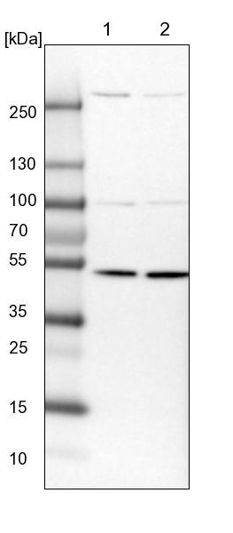
- Main image

- Experimental details
- Lane 1: NIH-3T3 cell lysate (Mouse embryonic fibroblast cells)Lane 2: NBT-II cell lysate (Rat Wistar bladder tumour cells)
- Submitted by
- Atlas Antibodies (provider)
- Main image
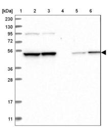
- Experimental details
- Lane 1: Marker [kDa] 230, 130, 95, 72, 56, 36, 28, 17, 11Lane 2: Human cell line RT-4Lane 3: Human cell line U-251MG spLane 4: Human plasma (IgG/HSA depleted)Lane 5: Human liver tissueLane 6: Human tonsil tissue

- Submitted by
- Atlas Antibodies (provider)
- Main image
- Experimental details
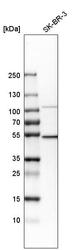
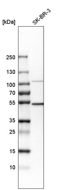
- Western blot analysis in human cell line SK-BR-3.
- Submitted by
- Atlas Antibodies (provider)
- Main image
- Experimental details
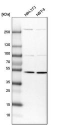
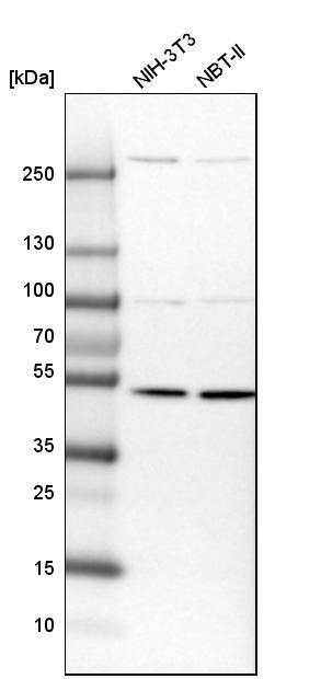
- Western blot analysis in mouse cell line NIH-3T3 and rat cell line NBT-II.
Supportive validation
- Submitted by
- Atlas Antibodies (provider)
- Main image
- Experimental details
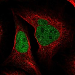
- Immunofluorescent staining of human cell line U-2 OS shows localization to nucleoplasm.
- Sample type
- HUMAN

Supportive validation
- Submitted by
- Atlas Antibodies (provider)
- Main image

- Experimental details
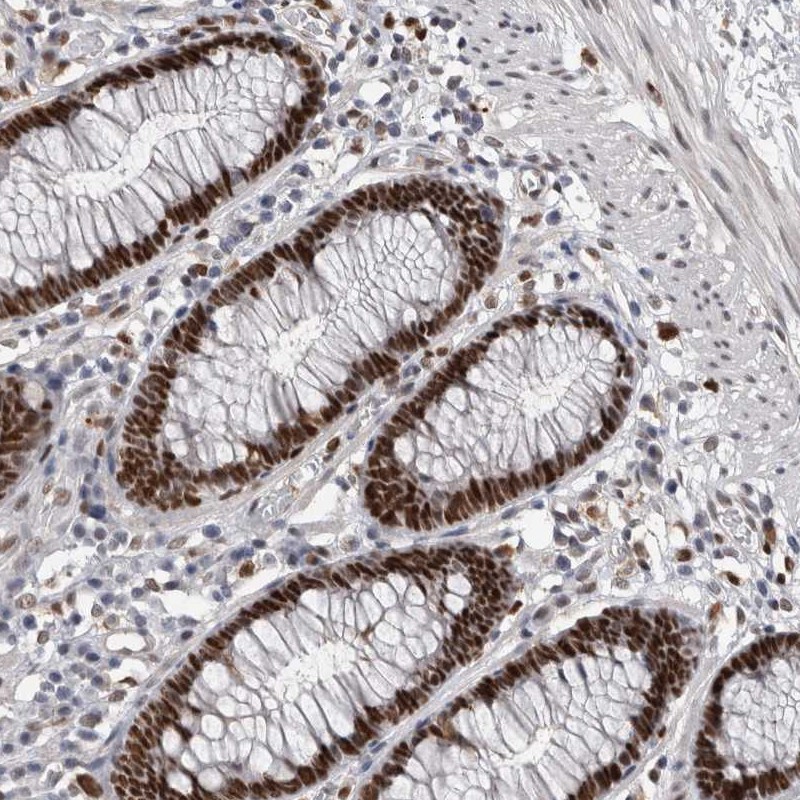
- Immunohistochemical staining of human colon shows strong nuclear positivity in glandular cells.
- Submitted by
- Atlas Antibodies (provider)
- Main image

- Experimental details
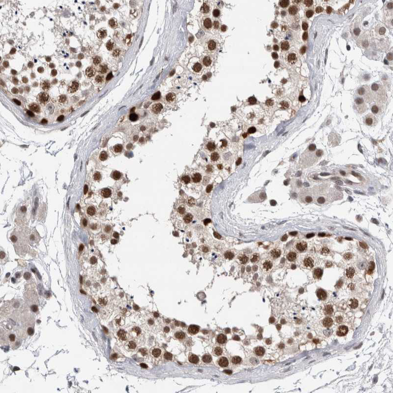
- Immunohistochemical staining of human testis shows moderate to strong nuclear positivity in cells in seminiferous ducts.
- Sample type
- HUMAN
- Submitted by
- Atlas Antibodies (provider)
- Main image

- Experimental details
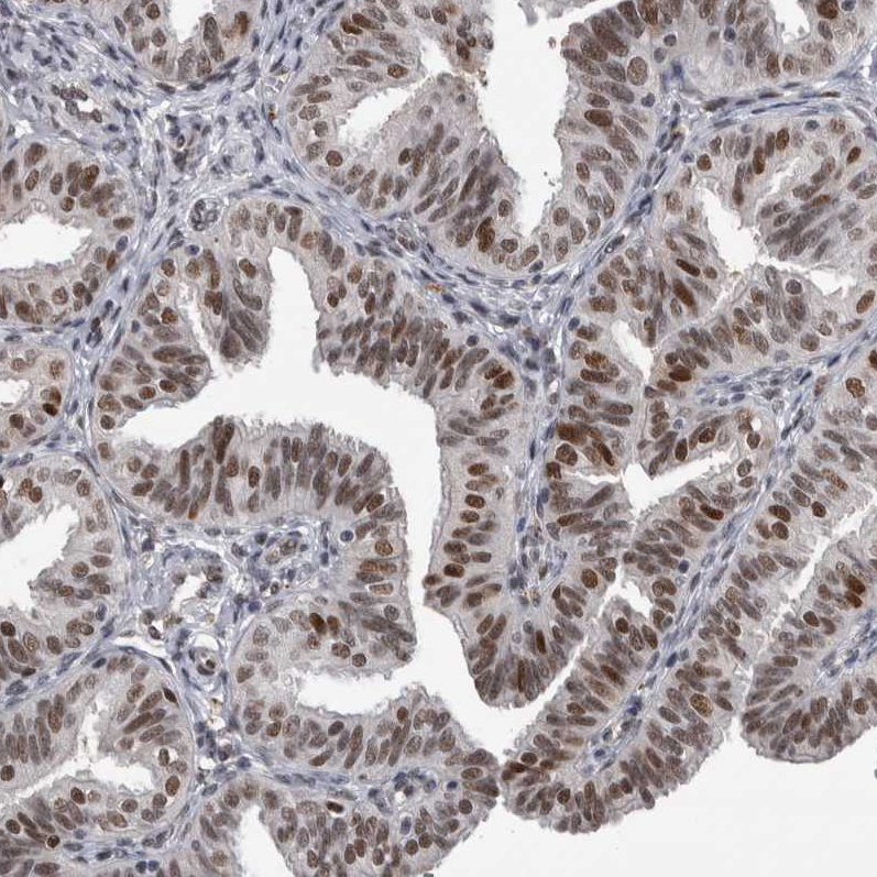
- Immunohistochemical staining of human Fallopian tube shows moderate nuclear positivity in glandular cells.
- Sample type
- HUMAN
- Submitted by
- Atlas Antibodies (provider)
- Main image
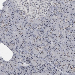
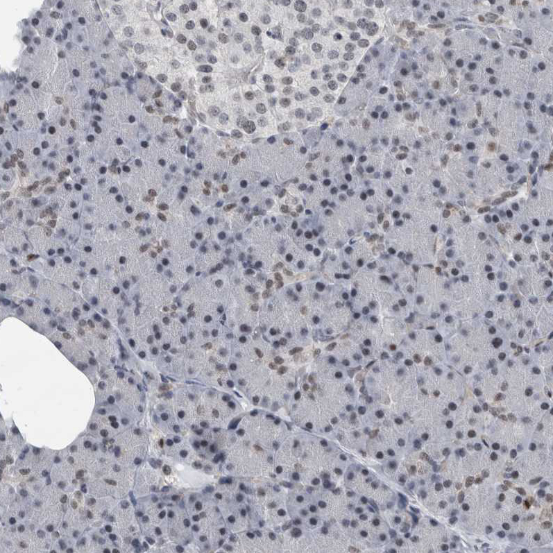
- Experimental details
- Immunohistochemical staining of human pancreas shows very weak positivity in exocrine glandular cells.
- Sample type
- HUMAN
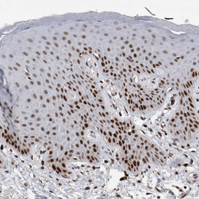
- Submitted by
- Atlas Antibodies (provider)
- Main image
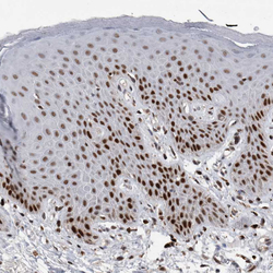
- Experimental details
- Immunohistochemical staining of human skin shows moderate to strong nuclear positivity in squamous epithelial cells.
- Sample type
- HUMAN