Explore
 Validate
 Learn
LearnABIN453704
antibody from antibodies-online
Targeting: CXCL8
3-10C, AMCF-I, b-ENAP, GCP-1, GCP1, IL-8, IL8, K60, LECT, LUCT, LYNAP, MDNCF, MONAP, NAF, NAP-1, NAP1, SCYB8, TSG-1
 Western blot
 ELISA
ELISAAntibody data
- Antibody Data
- Antigen structure
- References [2]
- Comments [0]
- Validations
- Western blot [1]
- Immunohistochemistry [1]
- Flow cytometry [1]
Submit
Validation data
Reference
Comment
Report error
- Product number
- ABIN453704 - Provider product page

- Provider
- antibodies-online
- Product name
- anti-Interleukin 8 (IL8) (C-Term) antibody
- Antibody type
- Polyclonal
- Antigen
- KLH conjugated synthetic peptide selected from the C-terminal region of human IL8
- Description
- Saturated Ammonium Sulfate (SAS) precipitation
- Reactivity
- Human
- Host
- Rabbit
- Epitope
- C-Term
- Vial size
- 0.4 mL
- Concentration
- 0.25 mg/mL
- Storage
- Store the antibody undiluted at 2-8°C for one month or (in aliquots) at -20°C for longer.
- Handling
- Avoid repeated freezing and thawing.
Submitted references Interleukin-8, a chemotactic and inflammatory cytokine.
Structure and functional expression of a human interleukin-8 receptor.
Baggiolini M, Clark-Lewis I
FEBS letters 1992 Jul 27;307(1):97-101
FEBS letters 1992 Jul 27;307(1):97-101
Structure and functional expression of a human interleukin-8 receptor.
Holmes WE, Lee J, Kuang WJ, Rice GC, Wood WI
Science (New York, N.Y.) 1991 Sep 13;253(5025):1278-80
Science (New York, N.Y.) 1991 Sep 13;253(5025):1278-80
No comments: Submit comment
Supportive validation
- Submitted by
- antibodies-online (provider)
- Main image
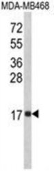
- Experimental details
- Western blot analysis of IL8 Antibody (C-term) (AP18073PU-N) in MDA-MB468 cell line lysates (35ug/lane). IL8 (arrow) was detected using the purified Pab.

Supportive validation
- Submitted by
- antibodies-online (provider)
- Main image
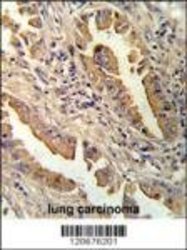
- Experimental details
- Formalin-fixed and paraffin-embedded human lung carcinoma reacated with IL8 antibody which was peroxidase-conjugated to the secondary antibody, followed by DAB staining.

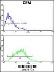
Supportive validation
- Submitted by
- antibodies-online (provider)
- Main image
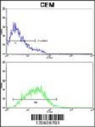
- Experimental details
- IL8 Antibody flow cytometry analysis of CEM cells (bottom) compared to negative control cells (top). FITC-conjugated goat-anti-rabbit secondary antibodies were employed for analysis.