 Explore
 Validate
 Learn
Learn Western blot
Antibody data
- Antibody Data
- Antigen structure
- References [0]
- Comments [0]
- Validations
- Western blot [1]
- Immunohistochemistry [1]
Submit
Validation data
Reference
Comment
Report error
- Product number
- AF8508 - Provider product page

- Provider
- R&D Systems
- Product name
- Human GIT1 Antibody
- Antibody type
- Polyclonal
- Description
- Immunogen affinity purified. Detects human GIT1 in direct ELISAs and Western blots.
- Reactivity
- Human
- Host
- Rabbit
- Conjugate
- Unconjugated
- Antigen sequence
Q9Y2X7- Isotype
- IgG
- Vial size
- 100 ug
- Storage
- Use a manual defrost freezer and avoid repeated freeze-thaw cycles. 12 months from date of receipt, -20 to -70 °C as supplied. 1 month, 2 to 8 °C under sterile conditions after reconstitution. 6 months, -20 to -70 °C under sterile conditions after reconstitution.
No comments: Submit comment
Supportive validation
- Submitted by
- R&D Systems (provider)
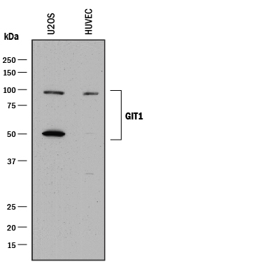
- Main image
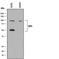
- Experimental details
- Detection of Human GIT1 by Western Blot. Western blot shows lysates of U2OS human osteosarcoma cell line and HUVEC human umbilical vein endothelial cells. PVDF membrane was probed with 0.025 µg/mL of Rabbit Anti-Human GIT1 Antigen Affinity-purified Polyclonal Antibody (Catalog # AF8508) followed by HRP-conjugated Anti-Rabbit IgG Secondary Antibody (Catalog # HAF008). Specific bands were detected for GIT1 at approximately 95 & 50 kDa (as indicated). This experiment was conducted under reducing conditions and using Immunoblot Buffer Group 1.
Supportive validation
- Submitted by
- R&D Systems (provider)
- Main image
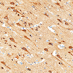
- Experimental details
- GIT1 in Human Brain. GIT1 was detected in immersion fixed paraffin-embedded sections of human brain (cortex) using Rabbit Anti-Human GIT1 Antigen Affinity-purified Polyclonal Antibody (Catalog # AF8508) at 0.3 µg/mL overnight at 4 °C. Tissue was stained using the Anti-Rabbit HRP-DAB Cell & Tissue Staining Kit (brown; Catalog # CTS005) and counterstained with hematoxylin (blue). Specific staining was localized to neurons. View our protocol for Chromogenic IHC Staining of Paraffin-embedded Tissue Sections.
