 Explore
 Validate
 Learn
Learn Western blot
Antibody data
- Antibody Data
- Antigen structure
- References [0]
- Comments [0]
- Validations
- Western blot [1]
- ELISA [1]
- Immunohistochemistry [1]
- Flow cytometry [1]
Submit
Validation data
Reference
Comment
Report error
- Product number
- MAB8508 - Provider product page

- Provider
- R&D Systems
- Product name
- Human/Mouse/Rat GIT1 Antibody
- Antibody type
- Monoclonal
- Description
- Protein A or G purified from hybridoma culture supernatant. Detects human GIT1 in direct ELISAs and human, mouse, and rat GIT1 in Western blots. In sandwich immunoassays, this antibody is specific for human GIT1 when paired with the suggested detection antibody.
- Reactivity
- Human, Mouse, Rat
- Host
- Mouse
- Conjugate
- Unconjugated
- Antigen sequence
Q9Y2X7- Isotype
- IgG
- Antibody clone number
- 924640
- Vial size
- 100 ug
- Storage
- Use a manual defrost freezer and avoid repeated freeze-thaw cycles. 12 months from date of receipt, -20 to -70 °C as supplied. 1 month, 2 to 8 °C under sterile conditions after reconstitution. 6 months, -20 to -70 °C under sterile conditions after reconstitution.
No comments: Submit comment
Supportive validation
- Submitted by
- R&D Systems (provider)
- Main image
- Experimental details
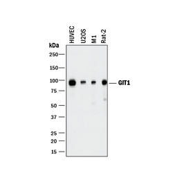
- Detection of Human, Mouse, and Rat GIT1 by Western Blot. Western blot shows lysates of HUVEC human umbilical vein endothelial cells, U2OS human osteosarcoma cell line, M1 mouse myeloid leukemia cell line, and Rat-2 rat embryonic fibroblast cell line. PVDF membrane was probed with 2 µg/mL of Mouse Anti-Human GIT1 Monoclonal Antibody (Catalog # MAB8508) followed by HRP-conjugated Anti-Mouse IgG Secondary Antibody (Catalog # HAF018). A specific band was detected for GIT1 at approximately 95 kDa (as indicated). This experiment was conducted under reducing conditions and using Immunoblot Buffer Group 1.

Supportive validation
- Submitted by
- R&D Systems (provider)
- Main image

- Experimental details
- Human GIT1 ELISA Standard Curve. Recombinant Human GIT1 protein was serially diluted 2-fold and captured by Mouse Anti-Human/Mouse/Rat GIT1 Monoclonal Antibody (Catalog # MAB8508) coated on a Clear Polystyrene Microplate (Catalog # DY990). Mouse Anti-Human GIT1 Monoclonal Antibody (Catalog # MAB85081) was biotinylated and incubated with the protein captured on the plate. Detection of the standard curve was achieved by incubating Streptavidin-HRP (Catalog # DY998) followed by Substrate Solution (Catalog # DY999) and stopping the enzymatic reaction with Stop Solution (Catalog # DY994).

Supportive validation
- Submitted by
- R&D Systems (provider)
- Main image
- Experimental details
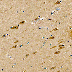
- GIT1 in Human Brain. GIT1 was detected in immersion fixed paraffin-embedded sections of human brain (hippocampus) using Mouse Anti-Human GIT1 Monoclonal Antibody (Catalog # MAB8508) at 15 µg/mL overnight at 4 °C. Tissue was stained using the Anti-Mouse HRP-DAB Cell & Tissue Staining Kit (brown; Catalog # CTS002) and counterstained with hematoxylin (blue). Specific staining was localized to neurons. View our protocol for Chromogenic IHC Staining of Paraffin-embedded Tissue Sections.

Supportive validation
- Submitted by
- R&D Systems (provider)
- Main image

- Experimental details
- Detection of GIT1 in SH-SY5Y Human Cell line by Flow Cytometry. SH-SY5Y human neuroblastoma cell line was stained with Mouse Anti-Human GIT1 Monoclonal Antibody (Catalog # MAB8508, filled histogram) or isotype control antibody (Catalog # MAB0041, open histogram), followed by Allophycocyanin-conjugated Anti-Mouse IgG Secondary Antibody (Catalog # F0101B). To facilitate intracellular staining, cells were fixed with Flow Cytometry Fixation Buffer (Catalog # FC004) and permeabilized with Flow Cytometry Permeabilization/Wash Buffer I (Catalog # FC005).
