Explore
 Validate
 Learn
Learn Western blot
Antibody data
- Antibody Data
- Antigen structure
- References [3]
- Comments [0]
- Validations
- Western blot [6]
- Immunocytochemistry [1]
Submit
Validation data
Reference
Comment
Report error
- Product number
- GTX106209 - Provider product page

- Provider
- GeneTex
- Proper citation
- GeneTex Cat#GTX106209, RRID:AB_1950203
- Product name
- Autotaxin antibody
- Antibody type
- Polyclonal
- Reactivity
- Human, Mouse, Rat
- Host
- Rabbit
Submitted references High Glucose Induces VEGF-C Expression via the LPA1/3-Akt-ROS-LEDGF Signaling Axis in Human Prostate Cancer PC-3 Cells.
Activation of Adenylyl Cyclase Causes Stimulation of Adenosine Receptors.
Association of autotaxin and lysophosphatidic acid receptor 3 with aggressiveness of human breast carcinoma.
Huang YL, Lin YC, Lin CC, Chen WM, Chen BPC, Lee H
Cellular physiology and biochemistry : international journal of experimental cellular physiology, biochemistry, and pharmacology 2018;50(2):597-611
Cellular physiology and biochemistry : international journal of experimental cellular physiology, biochemistry, and pharmacology 2018;50(2):597-611
Activation of Adenylyl Cyclase Causes Stimulation of Adenosine Receptors.
Pleli T, Mondorf A, Ferreiros N, Thomas D, Dvorak K, Biondi RM, Heringdorf DMZ, Zeuzem S, Geisslinger G, Zimmermann H, Waidmann O, Piiper A
Cellular physiology and biochemistry : international journal of experimental cellular physiology, biochemistry, and pharmacology 2018;45(6):2516-2528
Cellular physiology and biochemistry : international journal of experimental cellular physiology, biochemistry, and pharmacology 2018;45(6):2516-2528
Association of autotaxin and lysophosphatidic acid receptor 3 with aggressiveness of human breast carcinoma.
Popnikolov NK, Dalwadi BH, Thomas JD, Johannes GJ, Imagawa WT
Tumour biology : the journal of the International Society for Oncodevelopmental Biology and Medicine 2012 Dec;33(6):2237-43
Tumour biology : the journal of the International Society for Oncodevelopmental Biology and Medicine 2012 Dec;33(6):2237-43
No comments: Submit comment
Supportive validation
- Submitted by
- GeneTex (provider)
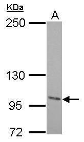
- Main image

- Experimental details
- Autotaxin antibody detects ENPP2 protein by western blot analysis.A. 30 ?g U87-MG whole cell lysate/extract5% SDS-PAGEAutotaxin antibody (GTX106209) dilution: 1:1000 The HRP-conjugated anti-rabbit IgG antibody (GTX213110-01) was used to detect the primary antibody.
- Submitted by
- GeneTex (provider)
- Main image
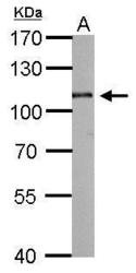
- Experimental details
- Autotaxin antibody detects ENPP2 protein by western blot analysis.A. 50 ?g mouse brain lysate/extract 7.5% SDS-PAGEAutotaxin antibody (GTX106209) dilution: 1:1000 The HRP-conjugated anti-rabbit IgG antibody (GTX213110-01) was used to detect the primary antibody.

- Submitted by
- GeneTex (provider)
- Main image

- Experimental details
- Autotaxin antibody detects ENPP2 protein by western blot analysis.A. 30 ?g Rat-2 whole cell lysate/extract5% SDS-PAGEAutotaxin antibody (GTX106209) dilution: 1:1000 The HRP-conjugated anti-rabbit IgG antibody (GTX213110-01) was used to detect the primary antibody.

- Submitted by
- GeneTex (provider)
- Main image

- Experimental details
- Various tissue extracts (30 ?g) were separated by 7.5% SDS-PAGE, and the membrane was blotted with Autotaxin antibody (GTX106209) diluted at 1:1000. The HRP-conjugated anti-rabbit IgG antibody (GTX213110-01) was used to detect the primary antibody.

- Submitted by
- GeneTex (provider)
- Main image

- Experimental details
- Human cerebrospinal fluid (30 ?g) was separated by 7.5% SDS-PAGE, and the membrane was blotted with Autotaxin antibody (GTX106209) diluted at 1:1000. The HRP-conjugated anti-rabbit IgG antibody (GTX213110-01) was used to detect the primary antibody.

- Submitted by
- GeneTex (provider)
- Main image

- Experimental details
- Human cerebrospinal fluid (15 ?g) was separated by 7.5% SDS-PAGE, and the membrane was blotted with Autotaxin antibody (GTX106209) diluted at 1:1000. The HRP-conjugated anti-rabbit IgG antibody (GTX213110-01) was used to detect the primary antibody.

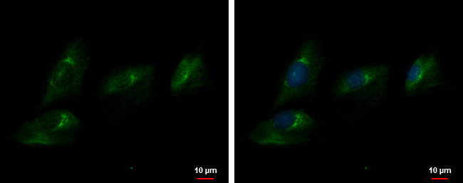
Supportive validation
- Submitted by
- GeneTex (provider)
- Main image

- Experimental details
- Autotaxin antibody detects Autotaxin protein at cytoplasm by immunofluorescent analysis.Sample: SK-N-SH cells were fixed in 4% paraformaldehyde at RT for 15 min.Green: Autotaxin protein stained by Autotaxin antibody (GTX106209) diluted at 1:500.Blue: Hoechst 33342 staining.