 Explore
 Validate
 Learn
Learn Western blot
 Immunocytochemistry
Antibody data
- Antibody Data
- Antigen structure
- References [1]
- Comments [0]
- Validations
- Western blot [2]
- Immunohistochemistry [7]
Submit
Validation data
Reference
Comment
Report error
- Product number
- NBP1-82563 - Provider product page

- Provider
- Novus Biologicals
- Proper citation
- Novus Cat#NBP1-82563, RRID:AB_11032030
- Product name
- Rabbit Polyclonal Njmu-R1 Antibody
- Antibody type
- Polyclonal
- Description
- Immunogen affinity purified. Specificity of human, mouse Njmu-R1 antibody verified on a Protein Array containing target protein plus 383 other non-specific proteins.
- Reactivity
- Human, Mouse
- Host
- Rabbit
- Isotype
- IgG
- Vial size
- 0.1 ml
- Storage
- Store at 4C short term. Aliquot and store at -20C long term. Avoid freeze-thaw cycles.
Submitted references Variance decomposition of protein profiles from antibody arrays using a longitudinal twin model.
Kato BS, Nicholson G, Neiman M, Rantalainen M, Holmes CC, Barrett A, Uhlén M, Nilsson P, Spector TD, Schwenk JM
Proteome science 2011 Nov 17;9:73
Proteome science 2011 Nov 17;9:73
No comments: Submit comment
Supportive validation
- Submitted by
- Novus Biologicals (provider)
- Main image

- Experimental details
- Western Blot: Njmu-R1 Antibody [NBP1-82563] - Lane 1: Marker [kDa] 250, 130, 100, 70, 55, 35, 25, 15, 10Lane 2: Mouse Cerebral Cortex tissue

- Submitted by
- Novus Biologicals (provider)
- Main image

- Experimental details
- Western Blot: Njmu-R1 Antibody [NBP1-82563] - Lane 1: Marker [kDa] 230, 110, 82, 49, 32, 26, 18Lane 2: Human cell line RT-4

Supportive validation
- Submitted by
- Novus Biologicals (provider)
- Main image

- Experimental details
- Immunohistochemistry-Paraffin: Njmu-R1 Antibody [NBP1-82563] - Staining of mouse somatosensory cortex shows immunoreactivity in cell bodies and dendrites of pyramidal neurons.
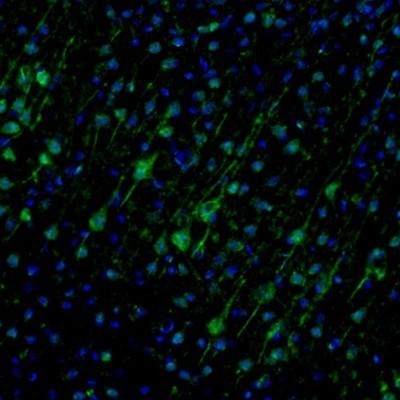
- Submitted by
- Novus Biologicals (provider)
- Main image

- Experimental details
- Immunohistochemistry-Paraffin: Njmu-R1 Antibody [NBP1-82563] - Staining of mouse olfactory bulb shows positivity in dendrites and cell bodies of mitral cells.

- Submitted by
- Novus Biologicals (provider)
- Main image

- Experimental details
- Immunohistochemistry-Paraffin: Njmu-R1 Antibody [NBP1-82563] - Staining of mouse piriform cortex shows staining in neuronal cell bodies and dendrites.
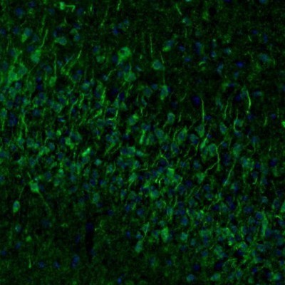
- Submitted by
- Novus Biologicals (provider)
- Main image

- Experimental details
- Immunohistochemistry-Paraffin: Njmu-R1 Antibody [NBP1-82563] - Staining of mouse dentate gyrus shows staining in a subset of neurons in granular layer.

- Submitted by
- Novus Biologicals (provider)
- Main image

- Experimental details
- Immunohistochemistry-Paraffin: Njmu-R1 Antibody [NBP1-82563] - Staining of mouse medulla shows neuronal staining in the inferior olivary nucleus.

- Submitted by
- Novus Biologicals (provider)
- Main image

- Experimental details
- Immunohistochemistry-Paraffin: Njmu-R1 Antibody [NBP1-82563] - Staining of human cerebral cortex shows dot-like positivity.

- Submitted by
- Novus Biologicals (provider)
- Main image

- Experimental details
- Immunohistochemistry-Paraffin: Njmu-R1 Antibody [NBP1-82563] - Staining of human stomach shows strong cytoplasmic positivity in glandular cells.
