 Explore
 Validate
 Learn
Learn Western blot
 Immunoprecipitation
Antibody data
- Antibody Data
- Antigen structure
- References [1]
- Comments [0]
- Validations
- Western blot [4]
- Immunocytochemistry [2]
- Immunohistochemistry [1]
Submit
Validation data
Reference
Comment
Report error
- Product number
- PA1-32440 - Provider product page

- Provider
- Invitrogen Antibodies
- Product name
- DJ-1 Polyclonal Antibody
- Antibody type
- Polyclonal
- Antigen
- Synthetic peptide
- Description
- PA1-32440 detects DJ1 from mouse, human, rat, hamster and fish samples.
- Reactivity
- Human, Mouse, Rat, Bovine, Chicken/Avian, Hamster, Zebrafish
- Host
- Rabbit
- Isotype
- IgG
- Vial size
- 100 µL
- Concentration
- Conc. Not Determined
- Storage
- Store at 4°C short term. For long term storage, store at -20°C, avoiding freeze/thaw cycles.
Submitted references Identification of potential therapeutic compounds for Parkinson's disease using Drosophila and human cell models.
Sanz FJ, Solana-Manrique C, Muñoz-Soriano V, Calap-Quintana P, Moltó MD, Paricio N
Free radical biology & medicine 2017 Jul;108:683-691
Free radical biology & medicine 2017 Jul;108:683-691
No comments: Submit comment
Supportive validation
- Submitted by
- Invitrogen Antibodies (provider)
- Main image

- Experimental details
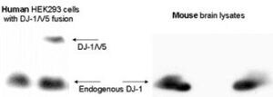
- Western Blot analysis of HEK293 cells overexpressin DJ-1-V5 tag protein and mouse brain lysates using DJ-1 Polyclonal Antibody (Product # PA1-32440).
- Submitted by
- Invitrogen Antibodies (provider)
- Main image

- Experimental details
- Western Blot analysis of DJ-1 was performed by loading human brain tissue lysate. Proteins were transferred to a membrane and probed with a DJ-1 Polyclonal Antibody (Product # PA1-32440).

- Submitted by
- Invitrogen Antibodies (provider)
- Main image

- Experimental details
- Knockdown of DJ-1 was achieved by transfecting HeLa cells with DJ-1 specific siRNAs (Silencer® select Product # s22304). Western blot analysis (Fig. a) was performed using whole cell extracts from the DJ-1 knockdown cells (lane 3), non-specific scrambled siRNA transfected cells (lane 2) and untransfected cells (lane 1). The blot was probed with DJ-1 Polyclonal Antibody (Product # PA1-32440, 1:1000 dilution) and Goat anti-Rabbit IgG (H+L), Superclonal™ Recombinant Secondary Antibody, HRP (Product # A27036, 0.25 µg/mL, 1:4000 dilution). Densitometric analysis of this western blot is shown in histogram (Fig. b). Decrease in signal upon siRNA mediated knock down confirms that antibody is specific to DJ-1.

- Submitted by
- Invitrogen Antibodies (provider)
- Main image

- Experimental details
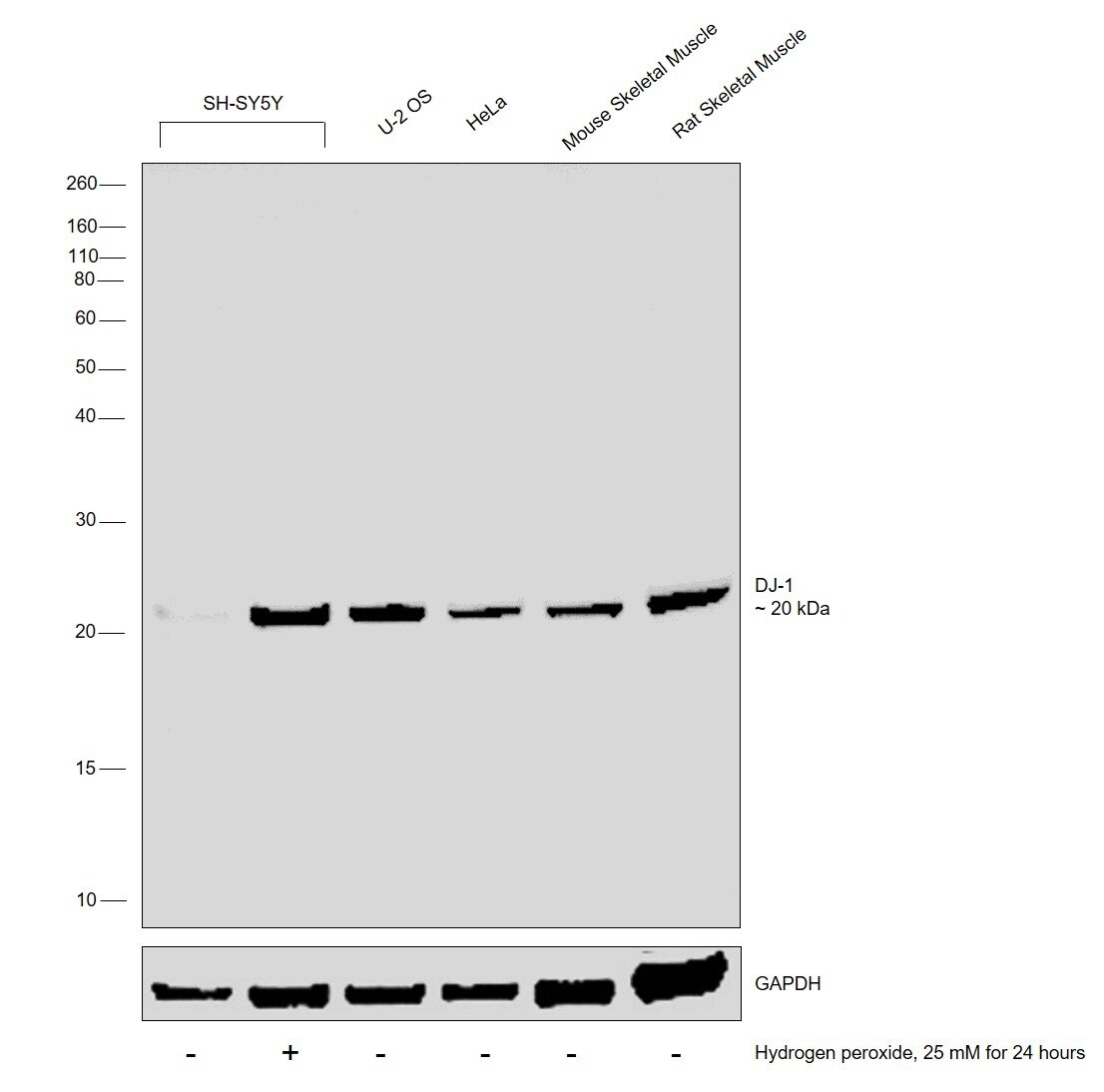
- Western blot was performed using DJ-1 Polyclonal Antibody (Product # PA1-32440) and a 20 kDa band corresponding to DJ-1 was observed in U-2 OS, HeLa, Mouse Skeletal Muscle and Rat Skeletal Muscle and increased upon Hydrogen peroxide treatment in SH-SY5Y. Whole cell extracts (30 µg lysate) of SH-SY5Y (Lane 1), SH-SY5Y treated with Hydrogen peroxide (25 mM for 24 hours) (Lane 2), U-2 OS (Lane 3), HeLa (Lane 4) and tissue extracts (30 µg lysate) of Mouse Skeletal Muscle (Lane 5) and Rat Skeletal Muscle (Lane 6) were electrophoresed using Novex® NuPAGE® 12 % Bis-Tris gel (Product # NP0342BOX). Resolved proteins were then transferred onto a nitrocellulose membrane (Product # IB23001) by iBlot® 2 Dry Blotting System (Product # IB21001). The blot was probed with the primary antibody (1:1000 dilution) and detected by chemiluminescence with Goat anti-Rabbit IgG (H+L), Superclonal™ Recombinant Secondary Antibody, HRP (Product # A27036, 1:4000 dilution) using the iBright FL 1000 (Product # A32752). Chemiluminescent detection was performed using Novex® ECL Chemiluminescent Substrate Reagent Kit (Product # WP20005).
Supportive validation
- Submitted by
- Invitrogen Antibodies (provider)
- Main image

- Experimental details
- Immunofluorescent analysis of DJ1 in human cortex using a DJ1 polyclonal antibody (Product # PA1-32440).

- Submitted by
- Invitrogen Antibodies (provider)
- Main image

- Experimental details
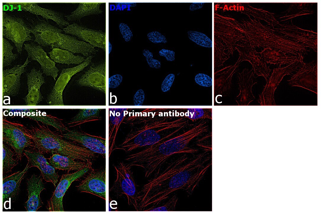
- Immunofluorescence analysis of DJ-1 was performed using 70% confluent log phase He-La cells. The cells were fixed with 4% Paraformaldehyde for 10 minutes, permeabilized with 0.1% Triton™ X-100 for 10 minutes, and blocked with 2% BSA for 1 hour at room temperature. The cells were labeled with DJ-1 Polyclonal Antibody (Product # PA1-32440) at 1:500 dilution in 0.1% BSA, incubated at 4 degree Celsius overnight and then labeled with Donkey anti-Rabbit IgG (H+L) Highly Cross-Adsorbed Secondary Antibody, Alexa Fluor Plus 488 (Product # A32790, 1:2000 dilution) for 45 minutes at room temperature (Panel a: Green). Nuclei (Panel b: Blue) were stained with SlowFade® Gold Antifade Mountant with DAPI (Product # S36938). F-actin (Panel c: Red) was stained with Rhodamine Phalloidin (Product # R415, 1:300). Panel d represents the merged image showing cytoplasmic and nuclear localization. Panel e represents control cells with no primary antibody to assess background. The images were captured at 60X magnification.
Supportive validation
- Submitted by
- Invitrogen Antibodies (provider)
- Main image

- Experimental details
- Immunohistochemistry (Paraffin) analysis of DJ-1 in human cortex tissue using DJ-1 Polyclonal Antibody (Product # PA1-32440).
