 Explore
 Validate
 Learn
Learn Western blot
Antibody data
- Antibody Data
- Antigen structure
- References [2]
- Comments [0]
- Validations
- Western blot [3]
- Immunocytochemistry [2]
Submit
Validation data
Reference
Comment
Report error
- Product number
- PA5-13404 - Provider product page

- Provider
- Invitrogen Antibodies
- Product name
- DJ-1 Polyclonal Antibody
- Antibody type
- Polyclonal
- Antigen
- Synthetic peptide
- Description
- This antibody is predicted to react with bovine and zebrafish based on sequence homology.
- Reactivity
- Human, Mouse
- Host
- Rabbit
- Isotype
- IgG
- Vial size
- 400 µL
- Concentration
- 2 mg/mL
- Storage
- Store at 4°C short term. For long term storage, store at -20°C, avoiding freeze/thaw cycles.
Submitted references The Mammalian Cap-Specific m(6)Am RNA Methyltransferase PCIF1 Regulates Transcript Levels in Mouse Tissues.
Decapping Enzyme NUDT12 Partners with BLMH for Cytoplasmic Surveillance of NAD-Capped RNAs.
Pandey RR, Delfino E, Homolka D, Roithova A, Chen KM, Li L, Franco G, Vågbø CB, Taillebourg E, Fauvarque MO, Pillai RS
Cell reports 2020 Aug 18;32(7):108038
Cell reports 2020 Aug 18;32(7):108038
Decapping Enzyme NUDT12 Partners with BLMH for Cytoplasmic Surveillance of NAD-Capped RNAs.
Wu H, Li L, Chen KM, Homolka D, Gos P, Fleury-Olela F, McCarthy AA, Pillai RS
Cell reports 2019 Dec 24;29(13):4422-4434.e13
Cell reports 2019 Dec 24;29(13):4422-4434.e13
No comments: Submit comment
Supportive validation
- Submitted by
- Invitrogen Antibodies (provider)
- Main image

- Experimental details
- Western blot analysis using a Park7 polyclonal antibody (Product # PA5-13404) in mouse brain, kidney, liver, NIH-3T3 and HeLa cell lysates (35 µg per lane).

- Submitted by
- Invitrogen Antibodies (provider)
- Main image
- Experimental details
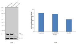
- Knockdown of DJ1 was achieved by transfecting SH-SY5Y cells with DJ1 specific siRNAs (Silencer® select Product # s22304, s22306). Western blot analysis (Fig. a) was performed using whole cell extracts from the DJ1 knockdown cells (lane 3), non-specific scrambled siRNA transfected cells (lane 2) and untransfected cells (lane 1). The blots were probed with DJ1 Polyclonal Antibody (Product # PA5-13404, 1:2000 dilution) and Goat anti-Rabbit IgG (H+L) Superclonal™ Secondary Antibody, HRP conjugate (Product # A27036, 0.25 µg/ml, 1:4000 dilution). Densitometric analysis of this western blot is shown in histogram (Fig. b). Decrease in signal upon siRNA mediated knock down confirms that antibody is specific to DJ1.

- Submitted by
- Invitrogen Antibodies (provider)
- Main image
- Experimental details
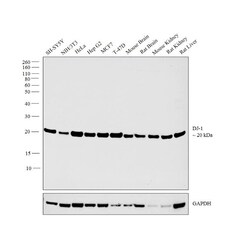
- Western blot analysis was performed on whole cell extracts (30 µg lysate) of SH-SY5Y (Lane 1), NIH/3T3 (Lane 2), HeLa (Lane 3), Hep G2 (Lane 4), MCF7 (Lane 5), T-47D (Lane 6), tissue extracts of Mouse Brain (Lane 7), Rat Brain (Lane 8), Mouse Kidney (Lane 9), Rat Kidney (Lane 10) and Rat Liver (Lane 11). The blot was probed with DJ-1 Polyclonal Antibody (Product # PA5-13404, 1:1000 dilution) and detected by chemiluminescence using Goat anti-Rabbit IgG (H+L) Superclonal™ Secondary Antibody, HRP conjugate (Product # A27036, 0.25 µg/ml, 1:4000 dilution). A band at 20 kDa corresponding to DJ-1 was observed across the cell lines and tissue tested.

Supportive validation
- Submitted by
- Invitrogen Antibodies (provider)
- Main image
- Experimental details
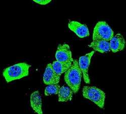
- Immunofluorescent analysis of HeLa cells using a Park7 polyclonal antibody (Product # PA5-13404) at a dilution of 1:10-50, followed by a fluor-conjugated goat anti-rabbit secondary antibody (green). Nuclei were stained with DAPI (blue).

- Submitted by
- Invitrogen Antibodies (provider)
- Main image
- Experimental details
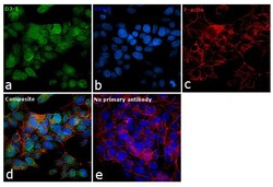
- Immunofluorescence analysis of DJ1 was performed using 70% confluent log phase SH-SY5Y cells. The cells were fixed with 4% paraformaldehyde for 10 minutes, permeabilized with 0.1% Triton™ X-100 for 15 minutes, and blocked with 1% BSA for 1 hour at room temperature. The cells were labeled with DJ1 Polyclonal Antibody (Product # PA5-13404) at 1:50 dilution in 0.1% BSA, incubated at 4 degree Celsius overnight and then labeled with Goat anti-Mouse IgG (H+L) Superclonal™ Secondary Antibody, Alexa Fluor® 488 conjugate (Product # A28175) at a dilution of 1:2000 for 45 minutes at room temperature (Panel a: green). Nuclei (Panel b: blue) were stained with ProLong™ Diamond Antifade Mountant with DAPI (Product # P36962). F-actin (Panel c: red) was stained with Rhodamine Phalloidin (Product # R415). Panel d represents the merged image showing nuclear and cytoplasmic localization. The images were captured at 60X magnification.
