Explore
 Validate
 Learn
Learn Western blot
 Immunoprecipitation
Antibody data
- Antibody Data
- Antigen structure
- References [1]
- Comments [0]
- Validations
- Western blot [3]
- Other assay [1]
Submit
Validation data
Reference
Comment
Report error
- Product number
- MA5-14870 - Provider product page

- Provider
- Invitrogen Antibodies
- Product name
- PIK3CA Monoclonal Antibody (H.843.0)
- Antibody type
- Monoclonal
- Antigen
- Synthetic peptide
- Description
- It is not recommended to aliquot this antibody.
- Reactivity
- Human, Mouse, Rat, Bovine
- Host
- Rabbit
- Isotype
- IgG
- Antibody clone number
- H.843.0
- Vial size
- 100 µL
- Concentration
- 121 µg/mL
- Storage
- -20°C
Submitted references Alterations of the renin angiotensin system in human end-stage heart failure before and after mechanical cardiac unloading by LVAD support.
Messmann R, Dietl A, Wagner S, Domenig O, Jungbauer C, Luchner A, Maier LS, Schopka S, Hirt S, Schmid C, Birner C
Molecular and cellular biochemistry 2020 Sep;472(1-2):79-94
Molecular and cellular biochemistry 2020 Sep;472(1-2):79-94
No comments: Submit comment
Supportive validation
- Submitted by
- Invitrogen Antibodies (provider)
- Main image

- Experimental details
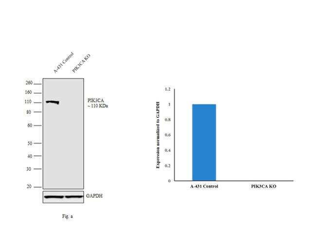
- Western blot analysis of PIK3CA (Fig. a) was performed by loading 30 µg of A-431 control (lane 1), A-431 PIK3CA knockout (lane 2) membrane extracts. PIK3CA was detected at 110 kDa using PIK3CA rabbit monoclonal antibody (Product # MA5-14870, 1:1000 dilution) and Goat anti-Rabbit IgG (H+L) Superclonal™ Secondary Antibody HRP conjugate (Product # A27036, 1:4000 dilution). Densitometric analysis of this western blot is shown in histogram (Fig. b). Loss of signal in CRISPR mediated knockout (KO) confirms that antibody is specific to PIK3CA.
- Submitted by
- Invitrogen Antibodies (provider)
- Main image
- Experimental details
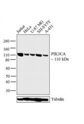
- Western blot analysis was performed on whole cell extracts (30 µg lysate) of Jurkat (Lane 1), HeLa (Lane 2), U-87 MG (Lane 3), SH-SY5Y (Lane 4) and A-431 (Lane 5). The blot was probed with Anti-PIK3CA Monoclonal Antibody (Product # MA5-14870, 1:1000 dilution) and detected by chemiluminescence using Goat anti-Rabbit IgG (H+L) Superclonal™ Secondary Antibody, HRP conjugate (Product # A27036, 0.25 µg/mL, 1:4000 dilution). A 110 kDa band corresponding to PIK3CA was observed across the cell lines tested.

- Submitted by
- Invitrogen Antibodies (provider)
- Main image

- Experimental details
- Western blot analysis was performed on whole cell extracts (30 µg lysate) of Jurkat (Lane 1), HeLa (Lane 2), U-87 MG (Lane 3), SH-SY5Y (Lane 4) and A-431 (Lane 5). The blot was probed with Anti-PIK3CA Monoclonal Antibody (Product # MA5-14870, 1:1000 dilution) and detected by chemiluminescence using Goat anti-Rabbit IgG (H+L) Superclonal™ Secondary Antibody, HRP conjugate (Product # A27036, 0.25 µg/mL, 1:4000 dilution). A 110 kDa band corresponding to PIK3CA was observed across the cell lines tested.

Supportive validation
- Submitted by
- Invitrogen Antibodies (provider)
- Main image
- Experimental details
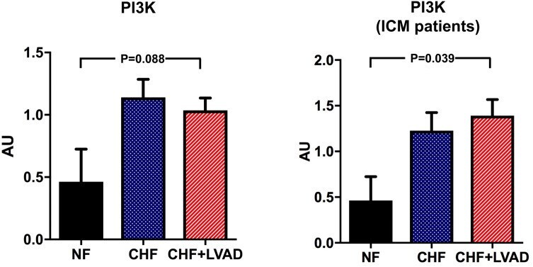
- Fig. 12 PI3K before (CHF) and after LVAD therapy (CHF+LVAD) as compared to non-failing ventricles (NF), left. The same comparisons in the subgroup of ICM patients, right ( n = 8). PI3K expression was determined by immunoblot (western blot) analysis and referred to a standard, respectively, whose densitometric value was set 1 by default. The value on the y -axis, therefore, reflects the percentage of each parameter's immunoblot band density in relation to this default value. AU arbitrary unit, CHF congestive heart failure, ICM ischemic cardiomyopathy, LVAD left ventricular assist device, NF non-failing myocardial tissue specimen