Explore
 Validate
 Learn
Learn Western blot
 Immunohistochemistry
Antibody data
- Antibody Data
- Antigen structure
- References [1]
- Comments [0]
- Validations
- Western blot [3]
- Immunohistochemistry [6]
Submit
Validation data
Reference
Comment
Report error
- Product number
- HPA004832 - Provider product page

- Provider
- Atlas Antibodies
- Proper citation
- Atlas Antibodies Cat#HPA004832, RRID:AB_1845044
- Product name
- Anti-ARPC1B
- Antibody type
- Polyclonal
- Reactivity
- Human, Mouse, Rat
- Host
- Rabbit
- Conjugate
- Unconjugated
- Antigen sequence
TVCLADADKKMAVATLASETLPLLALTFITDNSLV
AAGHDCFPVLFTYDAAAGMLSFGGRLDVPKQSSQR
GLTARERFQNLDKKASSEGGTAAGAGLDSLHKNSV
SQISVLSGGKAKCSQFCTTGMDGGMSIWDVKSLES
A- Isotype
- IgG
- Vial size
- 100 µl
- Storage
- Store at +4°C for short term storage. Long time storage is recommended at -20°C.
Submitted references Isoform diversity in the Arp2/3 complex determines actin filament dynamics
Abella J, Galloni C, Pernier J, Barry D, Kjær S, Carlier M, Way M
Nature Cell Biology 2015 December;18(1):76-86
Nature Cell Biology 2015 December;18(1):76-86
No comments: Submit comment
Supportive validation
Supportive validation
- Submitted by
- Atlas Antibodies (provider)
- Enhanced method
- Orthogonal validation
- Main image

- Experimental details
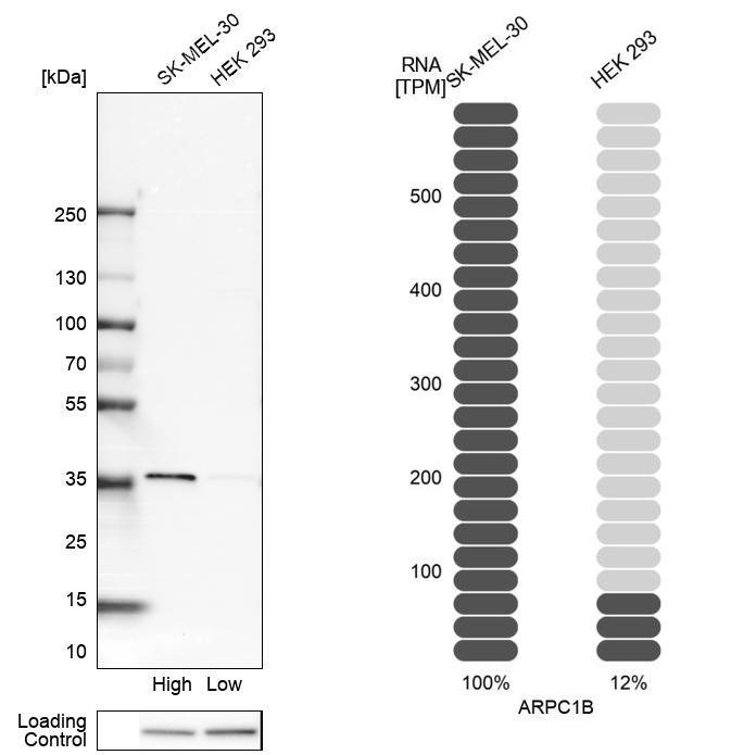
- Western blot analysis in human cell lines SK-MEL-30 and HEK293 using Anti-ARPC1B antibody. Corresponding ARPC1B RNA-seq data are presented for the same cell lines. Loading control: Anti-HDAC1.
Supportive validation
- Submitted by
- Atlas Antibodies (provider)
- Main image
- Experimental details
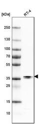
- Western blot analysis in human cell line RT-4.
- Sample type
- HUMAN

- Submitted by
- Atlas Antibodies (provider)
- Main image
- Experimental details
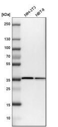
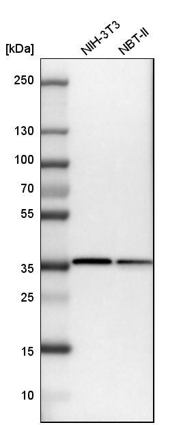
- Western blot analysis in mouse cell line NIH-3T3 and rat cell line NBT-II.
Enhanced validation
Supportive validation
- Submitted by
- Atlas Antibodies (provider)
- Enhanced method
- Orthogonal validation
- Main image

- Experimental details
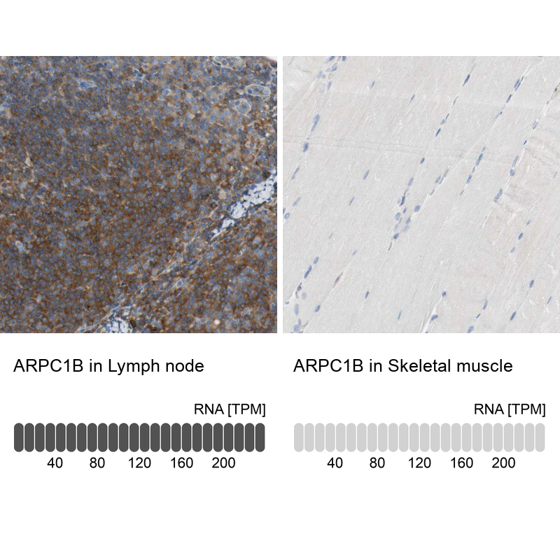
- Immunohistochemistry analysis in human lymph node and skeletal muscle tissues using HPA004832 antibody. Corresponding ARPC1B RNA-seq data are presented for the same tissues.
- Sample type
- HUMAN
Supportive validation
- Submitted by
- Atlas Antibodies (provider)
- Main image
- Experimental details
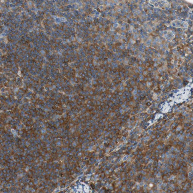
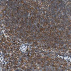
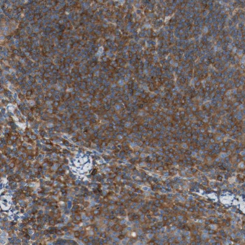
- Immunohistochemical staining of human lymph node shows strong cytoplasmic positivity in lymphoid cells outside reaction centra.
- Sample type
- HUMAN
- Submitted by
- Atlas Antibodies (provider)
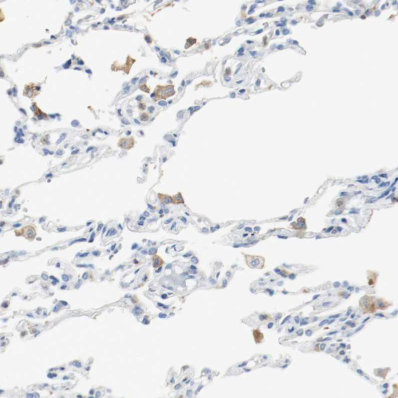
- Main image
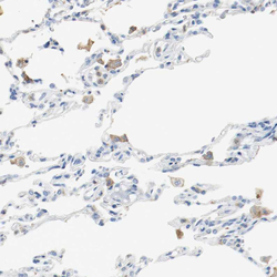
- Experimental details
- Immunohistochemical staining of human lung shows weak cytoplasmic positivity in macrophages.
- Sample type
- HUMAN
- Submitted by
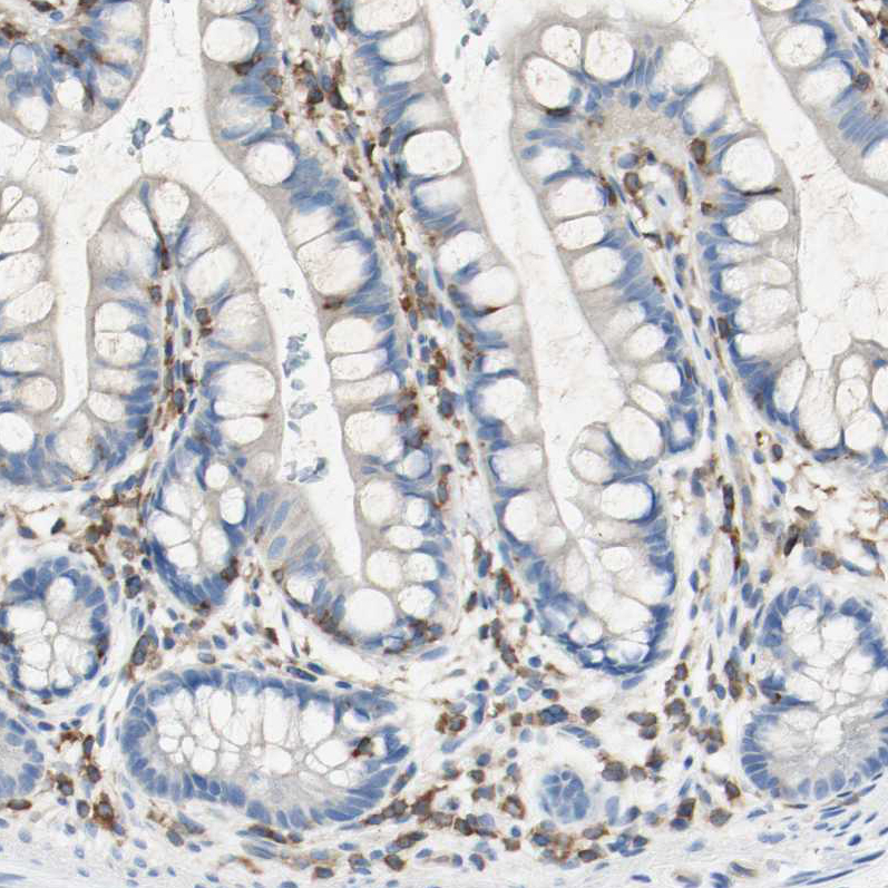
- Atlas Antibodies (provider)
- Main image
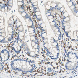
- Experimental details
- Immunohistochemical staining of human small intestine shows moderate cytoplasmic positivity in lymphoid cells.
- Sample type
- HUMAN
- Submitted by
- Atlas Antibodies (provider)
- Main image

- Experimental details
- Immunohistochemical staining of human skeletal muscle shows no positivity in myocytes as expected.
- Sample type
- HUMAN

- Submitted by
- Atlas Antibodies (provider)
- Main image

- Experimental details
- Immunohistochemical staining of human lymph node shows strong cytoplasmic positivity in germinal center cells.
- Sample type
- HUMAN