 Explore
 Validate
 Learn
Learn Immunocytochemistry
Antibody data
- Antibody Data
- Antigen structure
- References [7]
- Comments [0]
- Validations
- Immunocytochemistry [2]
- Immunohistochemistry [1]
- Flow cytometry [2]
Submit
Validation data
Reference
Comment
Report error
- Product number
- PA5-16770 - Provider product page

- Provider
- Invitrogen Antibodies
- Product name
- c-Kit Polyclonal Antibody
- Antibody type
- Polyclonal
- Antigen
- Synthetic peptide
- Description
- PA5-16770 targets CD117 in IHC (P) applications and shows reactivity with Canine, Rat and Human samples.
Submitted references Cardiac telocytes inhibit cardiac microvascular endothelial cell apoptosis through exosomal miRNA-21-5p-targeted cdip1 silencing to improve angiogenesis following myocardial infarction.
Mast Cells in the Auditory Periphery of Rodents.
Genetic diversity between mouse strains allows identification of the CC027/GeniUnc strain as an orally reactive model of peanut allergy.
Changes in the Neuronal Control of the Urinary Bladder in a Model of Radiation Cystitis.
SDF1 gradient associates with the distribution of c-Kit+ cardiac cells in the heart.
Phase 2 trial of dasatinib in target-selected patients with recurrent glioblastoma (RTOG 0627).
Wharton's jelly-derived mesenchymal stem cells promote myocardial regeneration and cardiac repair after miniswine acute myocardial infarction.
Liao Z, Chen Y, Duan C, Zhu K, Huang R, Zhao H, Hintze M, Pu Q, Yuan Z, Lv L, Chen H, Lai B, Feng S, Qi X, Cai D
Theranostics 2021;11(1):268-291
Theranostics 2021;11(1):268-291
Mast Cells in the Auditory Periphery of Rodents.
Szczepek AJ, Dudnik T, Karayay B, Sergeeva V, Olze H, Smorodchenko A
Brain sciences 2020 Oct 1;10(10)
Brain sciences 2020 Oct 1;10(10)
Genetic diversity between mouse strains allows identification of the CC027/GeniUnc strain as an orally reactive model of peanut allergy.
Orgel K, Smeekens JM, Ye P, Fotsch L, Guo R, Miller DR, Pardo-Manuel de Villena F, Burks AW, Ferris MT, Kulis MD
The Journal of allergy and clinical immunology 2019 Mar;143(3):1027-1037.e7
The Journal of allergy and clinical immunology 2019 Mar;143(3):1027-1037.e7
Changes in the Neuronal Control of the Urinary Bladder in a Model of Radiation Cystitis.
Giglio D, Podmolíková L, Tobin G
The Journal of pharmacology and experimental therapeutics 2018 May;365(2):327-335
The Journal of pharmacology and experimental therapeutics 2018 May;365(2):327-335
SDF1 gradient associates with the distribution of c-Kit+ cardiac cells in the heart.
Renko O, Tolonen AM, Rysä J, Magga J, Mustonen E, Ruskoaho H, Serpi R
Scientific reports 2018 Jan 18;8(1):1160
Scientific reports 2018 Jan 18;8(1):1160
Phase 2 trial of dasatinib in target-selected patients with recurrent glioblastoma (RTOG 0627).
Lassman AB, Pugh SL, Gilbert MR, Aldape KD, Geinoz S, Beumer JH, Christner SM, Komaki R, DeAngelis LM, Gaur R, Youssef E, Wagner H, Won M, Mehta MP
Neuro-oncology 2015 Jul;17(7):992-8
Neuro-oncology 2015 Jul;17(7):992-8
Wharton's jelly-derived mesenchymal stem cells promote myocardial regeneration and cardiac repair after miniswine acute myocardial infarction.
Zhang W, Liu XC, Yang L, Zhu DL, Zhang YD, Chen Y, Zhang HY
Coronary artery disease 2013 Nov;24(7):549-58
Coronary artery disease 2013 Nov;24(7):549-58
No comments: Submit comment
Supportive validation
- Submitted by
- Invitrogen Antibodies (provider)
- Main image
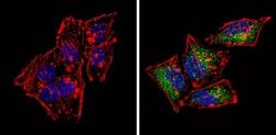
- Experimental details
- Immunofluorescent analysis of CD117 (green) showing staining in the nucleus and cytoplasm of Hela cells (right) compared to a negative control without primary antibody (left). Formalin-fixed cells were permeabilized with 0.1% Triton X-100 in TBS for 5-10 minutes and blocked with 3% BSA-PBS for 30 minutes at room temperature. Cells were probed with a CD117 polyclonal antibody (Product # PA5-16770) in 3% BSA-PBS at a dilution of 1:100 and incubated overnight at 4 ºC in a humidified chamber. Cells were washed with PBST and incubated with a DyLight-conjugated secondary antibody in PBS at room temperature in the dark. F-actin (red) was stained with a flourescent red phalloidin and nuclei (blue) were stained with Hoechst or DAPI. Images were taken at a magnification of 60x.

- Submitted by
- Invitrogen Antibodies (provider)
- Main image
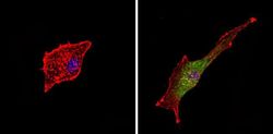
- Experimental details
- Immunofluorescent analysis of CD117 (green) showing staining in the nucleus and cytoplasm of U-87 MG cells (right) compared to a negative control without primary antibody (left). Formalin-fixed cells were permeabilized with 0.1% Triton X-100 in TBS for 5-10 minutes and blocked with 3% BSA-PBS for 30 minutes at room temperature. Cells were probed with a CD117 polyclonal antibody (Product # PA5-16770) in 3% BSA-PBS at a dilution of 1:100 and incubated overnight at 4 ºC in a humidified chamber. Cells were washed with PBST and incubated with a DyLight-conjugated secondary antibody in PBS at room temperature in the dark. F-actin (red) was stained with a flourescent red phalloidin and nuclei (blue) were stained with Hoechst or DAPI. Images were taken at a magnification of 60x.

Supportive validation
- Submitted by
- Invitrogen Antibodies (provider)
- Main image

- Experimental details
- Formalin-fixed, paraffin-embedded human stroma tumor stained with CD117/c-kit antibody using peroxidase-conjugate and AEC. Note staining of tumor cells.

Supportive validation
- Submitted by
- Invitrogen Antibodies (provider)
- Main image

- Experimental details
- Flow cytometry analysis of CD117 in C6 cells (green) compared to an isotype control (blue). Cells were harvested, adjusted to a concentration of 1-5x10^6 cells/ml, fixed with 2% paraformaldehyde and washed with PBS. Cells were blocked with a 2% solution of BSA-PBS for 30 min at room temperature and incubated with a CD117 polyclonal antibody (Product # PA5-16770) at a dilution of 2 ug/test for 60 min at room temperature. Cells were then incubated for 40 min at room temperature in the dark using a Dylight 488-conjugated secondary antibody and re-suspended in PBS for FACS analysis.

- Submitted by
- Invitrogen Antibodies (provider)
- Main image

- Experimental details
- Flow cytometry analysis of CD117 in PBMC cells (green) compared to an isotype control (blue). Human blood was collected, combined with a hydrophilic polysaccharide, centrifuged, transferred to a conical tube and washed with PBS. 50 ul of cell solution was added to each tube at a dilution of 2x10^7 cells/ml, followed by the addition of 50 ul of isotype control and primary antibody (Product # PA5-16770) at a dilution of 2 ug/test. Cells were incubated for 30 min at 4ºC and washed with a cell buffer, followed by incubation with a DyLight 488-conjugated secondary antibody for 30 min at 4ºC in the dark. FACS analysis was performed using 400 ul of cell buffer.
