Explore
 Validate
 Learn
LearnHPA021608
antibody from Atlas Antibodies
Targeting: HSD17B11
17-BETA-HSD11, 17-BETA-HSDXI, DHRS8, PAN1B, RetSDR2, SDR16C2
 Western blot
 Immunohistochemistry
Antibody data
- Antibody Data
- Antigen structure
- References [1]
- Comments [0]
- Validations
- Western blot [1]
- Immunocytochemistry [1]
- Immunohistochemistry [7]
Submit
Validation data
Reference
Comment
Report error
- Product number
- HPA021608 - Provider product page

- Provider
- Atlas Antibodies
- Proper citation
- Atlas Antibodies Cat#HPA021608, RRID:AB_1847660
- Product name
- Anti-HSD17B11
- Antibody type
- Polyclonal
- Reactivity
- Human
- Host
- Rabbit
- Conjugate
- Unconjugated
- Antigen sequence
FVNTGFIKNPSTSLGPTLEPEEVVNRLMHGILTEQ
KMIFIPSSIAFLTTLERILPERFLAVLKRKISVKF
DAVIGY- Isotype
- IgG
- Vial size
- 100 µl
- Storage
- Store at +4°C for short term storage. Long time storage is recommended at -20°C.
Submitted references RNA Deep Sequencing as a Tool for Selection of Cell Lines for Systematic Subcellular Localization of All Human Proteins
Danielsson F, Wiking M, Mahdessian D, Skogs M, Ait Blal H, Hjelmare M, Stadler C, Uhlén M, Lundberg E
Journal of Proteome Research 2013 January;12(1):299-307
Journal of Proteome Research 2013 January;12(1):299-307
No comments: Submit comment
Supportive validation
- Submitted by
- Atlas Antibodies (provider)
- Main image
- Experimental details
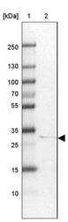
- Lane 1: Marker [kDa] 250, 130, 100, 70, 55, 35, 25, 15, 10Lane 2: Human cell line HepG2

Supportive validation
- Submitted by
- Atlas Antibodies (provider)
- Main image

- Experimental details
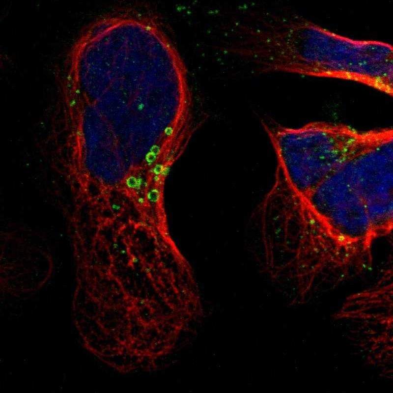
- Immunofluorescent staining of human cell line U-2 OS shows localization to lipid droplets.
- Sample type
- HUMAN
Enhanced validation
Supportive validation
- Submitted by
- Atlas Antibodies (provider)
- Enhanced method
- Orthogonal validation
- Main image

- Experimental details
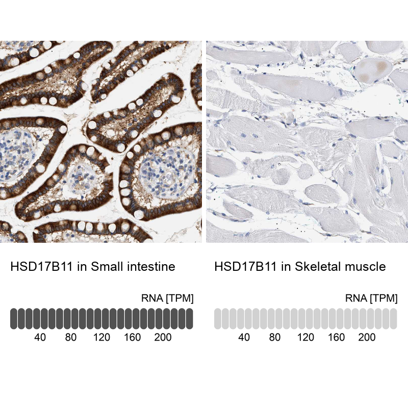
- Immunohistochemistry analysis in human small intestine and skeletal muscle tissues using HPA021608 antibody. Corresponding HSD17B11 RNA-seq data are presented for the same tissues.
- Sample type
- HUMAN
Supportive validation
- Submitted by
- Atlas Antibodies (provider)
- Main image

- Experimental details
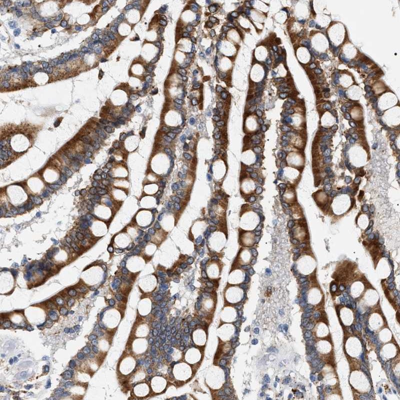
- Immunohistochemical staining of human colon shows strong cytoplasmic positivity in glandular cells.
- Submitted by
- Atlas Antibodies (provider)
- Main image
- Experimental details
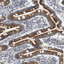
- Immunohistochemical staining of human duodenum shows moderate to strong cytoplasmic positivity in glandular cells.
- Sample type
- HUMAN
- Submitted by
- Atlas Antibodies (provider)
- Main image
- Experimental details
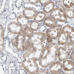
- Immunohistochemical staining of human kidney shows weak to moderate cytoplasmic positivity in cells in tubules.
- Sample type
- HUMAN

- Submitted by
- Atlas Antibodies (provider)
- Main image

- Experimental details
- Immunohistochemical staining of human colon shows moderate to strong cytoplasmic positivity in glandular cells.
- Sample type
- HUMAN
- Submitted by
- Atlas Antibodies (provider)
- Main image

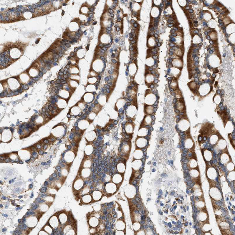
- Experimental details
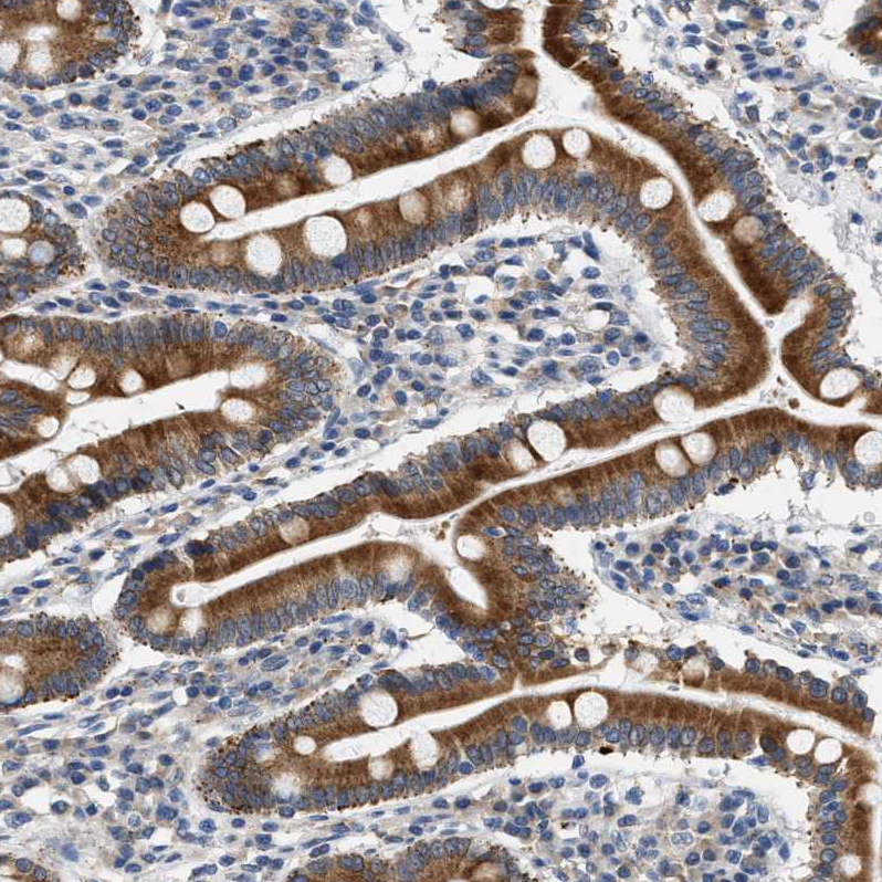
- Immunohistochemical staining of human small intestine shows strong cytoplasmic positivity in glandular cells.
- Sample type
- HUMAN

- Submitted by
- Atlas Antibodies (provider)
- Main image

- Experimental details
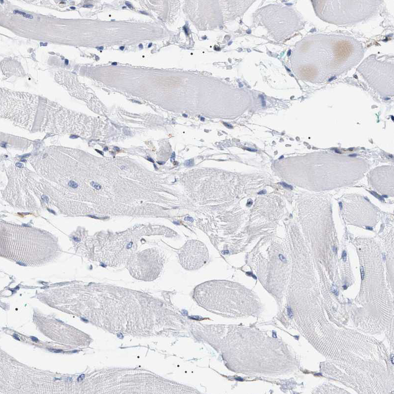
- Immunohistochemical staining of human skeletal muscle shows no positivity in myocytes as expected.
- Sample type
- HUMAN