Explore
 Validate
 Learn
Learn Western blot
Antibody data
- Antibody Data
- Antigen structure
- References [0]
- Comments [0]
- Validations
- Western blot [2]
- Immunocytochemistry [1]
- Immunohistochemistry [1]
- Flow cytometry [1]
Submit
Validation data
Reference
Comment
Report error
- Product number
- PA5-49447 - Provider product page

- Provider
- Invitrogen Antibodies
- Product name
- ITGA7 Polyclonal Antibody
- Antibody type
- Polyclonal
- Antigen
- Synthetic peptide
- Reactivity
- Human
- Host
- Rabbit
- Vial size
- 400 µL
- Concentration
- 0.5 mg/mL
- Storage
- -20° C, Avoid Freeze/Thaw Cycles
No comments: Submit comment
Supportive validation
- Submitted by
- Invitrogen Antibodies (provider)
- Main image

- Experimental details
- Western blot analysis of ITGA7 in A431 whole cell lysates (20 µg per lane). Samples were probed with an ITGA7 Antibody (N-term) (Product # PA5-49447) at a 1:4000 dilution. Secondary Goat Rabbit IgG, (H+L), Peroxidase conjugated at 1:10000 dilution. Predicted band size : 129 kDa. Blocking/Dilution buffer: 5% NFDM/TBST.

- Submitted by
- Invitrogen Antibodies (provider)
- Main image

- Experimental details
- Western blot analysis of ITGA7 in (Lane 1) Hela whole cell lysates (20 µg), (Lane 2) A431 whole cell lysates (20 µg), and (Lane 3) Jurkat whole cell lysates (20 µg). Samples were probed with a ITGA7 Antibody (N-term) (Product # PA5-49447) at 1:2000 dilution, followed by Secondary Goat Rabbit IgG, (H+L), Peroxidase conjugated at 1:10000 dilution. Predicted band size : 129 kDa. Blocking/Dilution buffer: 5% NFDM/TBST.

Supportive validation
- Submitted by
- Invitrogen Antibodies (provider)
- Main image

- Experimental details
- Immunofluorecent analysis of ITGA7 in human cervical epithelial adenocarcinoma HeLa cells. Cells were fixed, permeabilized and stained with ITGA7 polyclonal antibody (Product # PA5-49447) at a 1:25 dilution, followed by Dylight® 488-conjugated goat rabbit IgG (NK179883) secondary antibody at 1:200 dilution (green). Cytoplasmic actin is detected with Dylight® 554 Phalloidin (PD18466410) at 1:100 dilution (red). The nuclear counter stain is DAPI (blue).

Supportive validation
- Submitted by
- Invitrogen Antibodies (provider)
- Main image

- Experimental details
- Immunohistochemical analysis of ITGA7 in paraformaldehyde-fixed, paraffin-embedded human skeletal muscle tissue. Samples were incubated with a ITGA7 polyclonal antibody (Product # PA5-49447) at a 1:25 dilution for 1 hours at 37°C. A undiluted biotinylated goat polyvalent antibody was used as the secondary antibody.

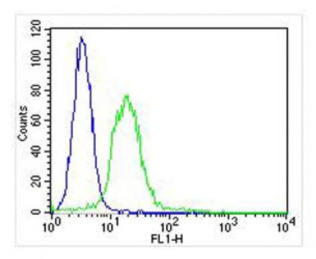
Supportive validation
- Submitted by
- Invitrogen Antibodies (provider)
- Main image

- Experimental details
- Flow cytometric analysis of ITGA7 in U-2 OS cells. Cells were fixed, permeabilized, and incubated with ITGA7 polyclonal antibody (Product # PA5-49447, green line) at a 1:25 dilution. After incubation with the primary antibody for 1 hour at 37°C, cells were incubated with a Goat-Rabbit IgG, DyLight® 488 Conjugated Highly Cross-Adsorbed antibody at 1/400 dilution for 40 min at 37°C. Isotype control antibody (blue line) was rabbit IgG (1µg/1x10^6 cells) used under the same conditions. Acquisition of >10,000 events was performed.