 Explore
 Validate
 Learn
LearnPAB10007
antibody from Abnova Corporation
Targeting: EGR1
AT225, G0S30, KROX-24, NGFI-A, TIS8, ZIF-268, ZNF225
 Western blot
Antibody data
- Antibody Data
- Antigen structure
- References [3]
- Comments [0]
- Validations
- Western blot [1]
- Immunohistochemistry [1]
Submit
Validation data
Reference
Comment
Report error
- Product number
- PAB10007 - Provider product page

- Provider
- Abnova Corporation
- Proper citation
- Abnova Corporation Cat#PAB10007, RRID:AB_1674714
- Product name
- EGR1 polyclonal antibody
- Antibody type
- Polyclonal
- Description
- Rabbit polyclonal antibody raised against synthetic peptide of EGR1.
- Storage
- Store at 4°C. For long term storage store at -20°C.Aliquot to avoid repeated freezing and thawing.
Submitted references A gene coding for a zinc finger protein is induced during 12-O-tetradecanoylphorbol-13-acetate-stimulated HL-60 cell differentiation.
Expression of a zinc finger gene in HTLV-I- and HTLV-II-transformed cells.
cDNA sequence of the human cellular early growth response gene Egr-1.
Shimizu N, Ohta M, Fujiwara C, Sagara J, Mochizuki N, Oda T, Utiyama H
Journal of biochemistry 1992 Feb;111(2):272-7
Journal of biochemistry 1992 Feb;111(2):272-7
Expression of a zinc finger gene in HTLV-I- and HTLV-II-transformed cells.
Wright JJ, Gunter KC, Mitsuya H, Irving SG, Kelly K, Siebenlist U
Science (New York, N.Y.) 1990 May 4;248(4955):588-91
Science (New York, N.Y.) 1990 May 4;248(4955):588-91
cDNA sequence of the human cellular early growth response gene Egr-1.
Suggs SV, Katzowitz JL, Tsai-Morris C, Sukhatme VP
Nucleic acids research 1990 Jul 25;18(14):4283
Nucleic acids research 1990 Jul 25;18(14):4283
No comments: Submit comment
Supportive validation
- Submitted by
- Abnova Corporation (provider)
- Main image

- Experimental details
- Western blot using EGR1 polyclonal antibody (Cat # PAB10007) shows detectionof a predominant band at ~58 KDa correspond-ing to EGR1 present in mouse embryonic fibroblast whole cell lysate (arrowhead).Approximately 35 ug of lysate was separated by 4-20% SDS-PAGE and transferred onto nitrocellulose.After blocking the membrane was probed with the primary antibody diluted to 1:1,500.Reaction occurred 2h at room temperature followed by washes and reaction with a 1 : 10,000 dilution of IRDye™800 conjugated Gt-a-Rabbit IgG [H&L] MX for 45 min at room temperature.

Supportive validation
- Submitted by
- Abnova Corporation (provider)
- Main image
- Experimental details
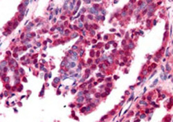
- Immunohistochemistry of EGR1 polyclonal antibody (Cat # PAB10007) was used at a 10 ug/mL to detect nuclear and cytoplasmic signal with low background staining in a variety of tissues including multi-human, multi-brain and multi-cancer slides.Within the multi-tumor block, the antibody showed variable levels of nuclear and cytoplasmic staining between individual tumors, with some tumors showing moderate staining.This image shows EGR1 staining of human ovarian carcinoma. Tissue was formalin-fixed and paraffin embedded.Personal Communication, Tina Roush, Life Span Biosciences, Seattle, WA.
- Validation comment
- Immunohistochemistry (Formalin/PFA-fixed paraffin-embedded sections)
